Femur: Difference between revisions
No edit summary |
Kim Jackson (talk | contribs) m (Text replacement - "[[Hip Fracture/Femoral Neck Fracture|" to "[[Femoral Neck Hip Fracture|") |
||
| (5 intermediate revisions by one other user not shown) | |||
| Line 6: | Line 6: | ||
== Introduction == | == Introduction == | ||
[[File:Femur bone.png|right|frameless|801x801px]] | [[File:Femur bone.png|right|frameless|801x801px]] | ||
The femur is the longest, heaviest, and strongest bone in the human body. | The femur is the longest, heaviest, and strongest [[bone]] in the human body. The main function of the femur is [[weight bearing]] and stability of [[Gait|gait.]] An essential component of the lower [[Closed Chain Exercise|kinetic chain]]. | ||
= | The robust shape of the femur provides many sturdy attachment points for the powerful [[:Category:Hip - Muscles|muscles of the hip]] and [[:Category:Knee - Muscles|knee]] that contribute to [[Walking - Muscles Used|walking]] and other propulsive movements.<ref name="Moore">Moore KL, Agur AM, Dalley AF. Essential Clinical Anatomy. 4th ed. Baltimore, MD: Lippincott Williams and Wilkins, 2011.</ref> | ||
== Femur: Three Divisions == | |||
The femur acts as the site of origin and attachment of many muscles and [[Functional Anatomy of the Hip-Bones and Ligaments|ligaments]], and can be divided into three parts; proximal, shaft and distal. <ref name=":0">Chang A, Breeland G, Hubbard JB. [https://www.ncbi.nlm.nih.gov/books/NBK532982/ Anatomy, Bony Pelvis and Lower Limb, Femur.] InStatPearls [Internet] 2019 Jul 3. StatPearls Publishing.Available from:https://www.ncbi.nlm.nih.gov/books/NBK532982/ (last accessed 16.6.2020)</ref> | |||
[[File:Femur front.png|right|frameless|939x939px]] | |||
'''Proximal Femur: C'''omposed of the head, neck, greater trochanter and lesser trochanter. The head of the femur articulates with the acetabulum of the [[pelvis]] to create the [[Hip Anatomy|hip joint]]. | |||
# Femoral head: Supported by the neck of the femur.Is a globular form rather more than a hemisphere. It is directed caudally, medially and anteriorly, known as the angle of inclination. Normal 120 degrees, abnormal angles; [[Coxa Vara / Coxa Valga|coxa vara]] less than 125 degrees; [[Coxa Vara / Coxa Valga|coxa valga]] greater than 125 degrees. Its surface is smooth and coated in [[cartilage]] except for an ovoid depression, fovea capitis femoris (a little below and behind the centre of the head). The fovea allows for the attachment of the ligament of head of the femur. | |||
# The bulbous femoral head is joined to the shaft of the femur by the femoral neck. If there is a [[Femoral Neck Hip Fracture|femoral neck fracture]], the blood supply through the ligament becomes crucial, and there is a risk of [[Avascular necrosis of the femoral head|avascular necrosis.]] | |||
# At the base of the neck are the medially oriented lesser trochanter and laterally placed greater trochanter. A rough line called the intertrochanteric line connects the greater and lesser trochanter on the anterior aspect of the femur, while the smoother intertrochanteric crest connects the trochanters posteriorly.<ref name="Moore" /><ref name="Neumann" /> | |||
'''Femoral Shaft''' | '''Femoral Shaft''' | ||
The femoral shaft is almost cylindrical in form, being slightly broader superiorly and slightly arched, giving it a convexity anteriorly and concavity posteriorly which has a prominent longitudinal ridge of bone, the linea aspera. A variety of muscles have their origins at and insert into the femoral shaft (all, apart from gluteus maximus and vastus intermedius, interact with the posterior surface of the bone).<ref name=":1">Teach me anatomy [https://teachmeanatomy.info/lower-limb/bones/femur/ Femur] Available from:https://teachmeanatomy.info/lower-limb/bones/femur/ (last accessed 16.6.2020)</ref> | |||
'''Distal Femur''' | '''Distal Femur''' | ||
| Line 35: | Line 28: | ||
== Blood Supply == | == Blood Supply == | ||
The femoral artery is the main blood supply to the lower extremity. | The femoral artery is the main blood supply to the lower extremity. The foveal artery comes off of the obturator artery that runs through the ligamentum teres femoris as supportive blood supply to the femoral head, but it is not the main source during adulthood. The perforating branches of the deep femoral artery supply the shaft and distal portion of the femur.<ref name=":0" />See also [[Functional Anatomy of the Hip - Neural and Vascular]] | ||
== Muscles == | == Muscles == | ||
[[File:Quadriceps muscle.jpg|right|frameless|450x450px]] | [[File:Quadriceps muscle.jpg|right|frameless|450x450px]] | ||
The thigh muscles are divided into the anterior, medial, and posterior and gluteal compartments. The femur sits within the anterior compartment. | The thigh muscles are divided into the anterior, medial, and posterior and gluteal compartments. The femur sits within the anterior compartment. | ||
# Anterior compartment is composed of | # Anterior compartment is composed of the [[Hip Flexors|hip flexors]] and [[Knee Extensors|knee extensors]]. Hip flexors include pectineus, iliopsoas, and sartorius muscle. | ||
# Medial compartment’s function is mainly leg adduction. | # Medial compartment’s function is mainly leg adduction. See [[Hip Adductors]] | ||
# Posterior compartment muscles are mainly hip extensors and knee flexors. | # Posterior compartment muscles are mainly hip extensors and knee flexors. [[Hip Extensors|Hip extensors]] [[Knee Flexors]] | ||
# Gluteal | # [[Gluteal Muscles|Gluteal]] muscles. 1.The superficial layer is composed of the gluteus maximus, medius, and minimus. Hip extension, abduction, and internal rotation is the superficial gluteal’s main function. 2. The deep layer is made up of the piriformis, obturator internus, quadratus femoris, and superior and inferior gemellus (deeper gluteal muscles help with external rotation of the hip)<ref name=":0" />. | ||
== Articulations == | == Articulations == | ||
# The femoral head of the proximal femur articulates with the acetabulum of the pelvis in which the femoral head acts at the ball and the acetabulum as the socket. Allows for movement at the [[Hip|hip]] in three [[Cardinal Planes and Axes of Movement|planes]]: flexion and extension in the sagittal plane, abduction and adduction in the frontal plane, and internal and external rotation in the horizontal plane.<ref name="Neumann">Neumann DA, Kinesiology of the musculoskeletal system: Foundations for rehabilitation. 2nd ed. St. Louis, MO: Mosby Elsevier, 2010. p520-71.</ref> | # The femoral head of the proximal femur articulates with the acetabulum of the pelvis in which the femoral head acts at the ball and the acetabulum as the socket. Allows for movement at the [[Hip|hip]] in three [[Cardinal Planes and Axes of Movement|planes]]: flexion and extension in the sagittal plane, abduction and adduction in the frontal plane, and internal and external rotation in the horizontal plane.<ref name="Neumann">Neumann DA, Kinesiology of the musculoskeletal system: Foundations for rehabilitation. 2nd ed. St. Louis, MO: Mosby Elsevier, 2010. p520-71.</ref> | ||
# Distally, the convex femoral condyles of the femur articulate with the condyles of the [[Tibia|tibia]], ie [[Knee|tibiofemoral joint]]. Movement at the tibiofemoral joint occurs in two planes: knee flexion and extension in the sagittal plane, and internal and external rotation in the horizontal plane.<ref name="Neumann" / | # Distally, the convex femoral condyles of the femur articulate with the condyles of the [[Tibia|tibia]], ie [[Knee|tibiofemoral joint]]. Movement at the tibiofemoral joint occurs in two planes: knee flexion and extension in the sagittal plane, and internal and external rotation in the horizontal plane.<ref name="Neumann" /> | ||
# The patellofemoral joint is formed by the articulation of the [[ | # The patellofemoral joint is formed by the articulation of the [[patella]] with the intercondylar/trochlear groove of the femur. During flexion and extension of the knee, the articular surfaces of the patella and femur perform a sliding movement.<ref name="Neumann" /> | ||
== Injuries and Conditions == | == Injuries and Conditions == | ||
[[File: | [[File:Femoral-shaft-fracture.jpeg|thumb|220x220px|Femoral Shaft Fracture]]See: | ||
<u>[[Femoral stress fracture|Femoral Stress fracture]]</u> | * [[Femoral Fractures|Femoral fractures]] | ||
* <u>[[Femoral stress fracture|Femoral Stress fracture]]</u> | |||
[[Patellofemoral Pain Syndrome|Patellofemoral pain syndrome]] .<ref name="Neumann" />.<ref>Fullem BW.Overuse lower extremity injuries in sport | * [[Patellofemoral Pain Syndrome|Patellofemoral pain syndrome]] .<ref name="Neumann" />.<ref>Fullem BW.Overuse lower extremity injuries in sport | ||
Clin Podiatr Med Surg. 2015 Apr;32(2):239-251. doi: 10.1016/j.cpm.2014.11.006. Epub 2014 Dec 30. Review. | Clin Podiatr Med Surg. 2015 Apr;32(2):239-251. doi: 10.1016/j.cpm.2014.11.006. Epub 2014 Dec 30. Review. | ||
PMID: 25804713</ref> | PMID: 25804713</ref> | ||
* [[Femoral Neck Fractures|Femoral Neck Fracture]] | |||
* [[Greater Trochanteric Pain Syndrome]] | |||
== References == | == References == | ||
<references /> | <references /> | ||
Latest revision as of 12:08, 19 December 2022
Original Editor - Daphne Jackson
Top Contributors - Lucinda hampton, Kim Jackson, Evan Thomas, Admin, Daphne Jackson, Joanne Garvey, Joao Costa and WikiSysop
Introduction[edit | edit source]
The femur is the longest, heaviest, and strongest bone in the human body. The main function of the femur is weight bearing and stability of gait. An essential component of the lower kinetic chain.
The robust shape of the femur provides many sturdy attachment points for the powerful muscles of the hip and knee that contribute to walking and other propulsive movements.[1]
Femur: Three Divisions[edit | edit source]
The femur acts as the site of origin and attachment of many muscles and ligaments, and can be divided into three parts; proximal, shaft and distal. [2]
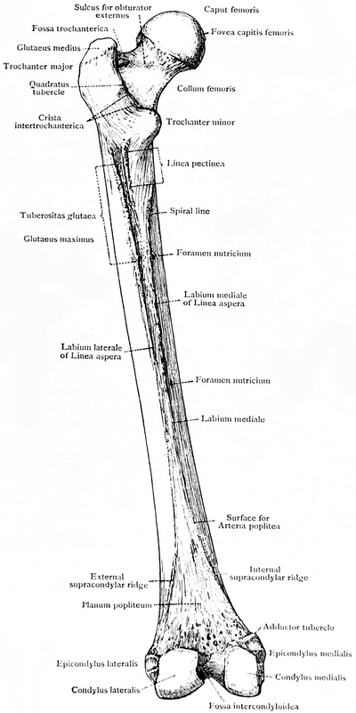
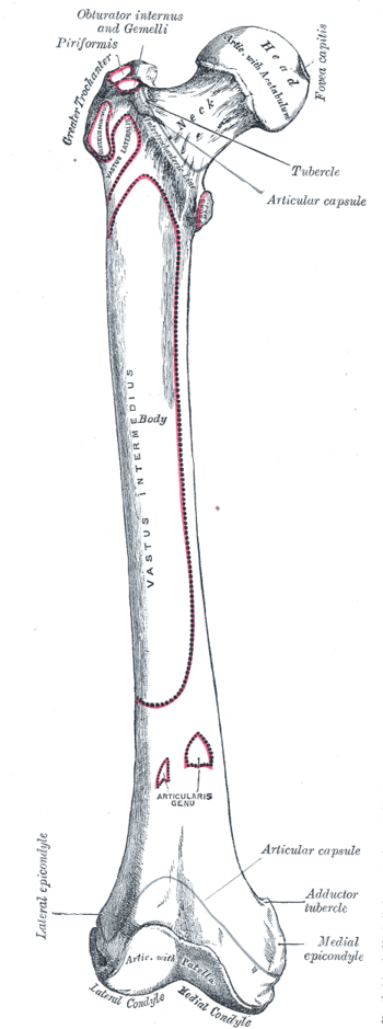
Proximal Femur: Composed of the head, neck, greater trochanter and lesser trochanter. The head of the femur articulates with the acetabulum of the pelvis to create the hip joint.
- Femoral head: Supported by the neck of the femur.Is a globular form rather more than a hemisphere. It is directed caudally, medially and anteriorly, known as the angle of inclination. Normal 120 degrees, abnormal angles; coxa vara less than 125 degrees; coxa valga greater than 125 degrees. Its surface is smooth and coated in cartilage except for an ovoid depression, fovea capitis femoris (a little below and behind the centre of the head). The fovea allows for the attachment of the ligament of head of the femur.
- The bulbous femoral head is joined to the shaft of the femur by the femoral neck. If there is a femoral neck fracture, the blood supply through the ligament becomes crucial, and there is a risk of avascular necrosis.
- At the base of the neck are the medially oriented lesser trochanter and laterally placed greater trochanter. A rough line called the intertrochanteric line connects the greater and lesser trochanter on the anterior aspect of the femur, while the smoother intertrochanteric crest connects the trochanters posteriorly.[1][3]
Femoral Shaft
The femoral shaft is almost cylindrical in form, being slightly broader superiorly and slightly arched, giving it a convexity anteriorly and concavity posteriorly which has a prominent longitudinal ridge of bone, the linea aspera. A variety of muscles have their origins at and insert into the femoral shaft (all, apart from gluteus maximus and vastus intermedius, interact with the posterior surface of the bone).[4]
Distal Femur
Prominent lateral and medial condyles are found at the distal end of the femur. Projecting from each condyle is an epicondyle that act as attachment sites for the collateral ligaments. The lateral and medial condyles are separated by the intercondylar notch.[3]
Blood Supply[edit | edit source]
The femoral artery is the main blood supply to the lower extremity. The foveal artery comes off of the obturator artery that runs through the ligamentum teres femoris as supportive blood supply to the femoral head, but it is not the main source during adulthood. The perforating branches of the deep femoral artery supply the shaft and distal portion of the femur.[2]See also Functional Anatomy of the Hip - Neural and Vascular
Muscles[edit | edit source]
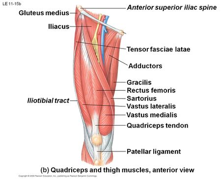
The thigh muscles are divided into the anterior, medial, and posterior and gluteal compartments. The femur sits within the anterior compartment.
- Anterior compartment is composed of the hip flexors and knee extensors. Hip flexors include pectineus, iliopsoas, and sartorius muscle.
- Medial compartment’s function is mainly leg adduction. See Hip Adductors
- Posterior compartment muscles are mainly hip extensors and knee flexors. Hip extensors Knee Flexors
- Gluteal muscles. 1.The superficial layer is composed of the gluteus maximus, medius, and minimus. Hip extension, abduction, and internal rotation is the superficial gluteal’s main function. 2. The deep layer is made up of the piriformis, obturator internus, quadratus femoris, and superior and inferior gemellus (deeper gluteal muscles help with external rotation of the hip)[2].
Articulations[edit | edit source]
- The femoral head of the proximal femur articulates with the acetabulum of the pelvis in which the femoral head acts at the ball and the acetabulum as the socket. Allows for movement at the hip in three planes: flexion and extension in the sagittal plane, abduction and adduction in the frontal plane, and internal and external rotation in the horizontal plane.[3]
- Distally, the convex femoral condyles of the femur articulate with the condyles of the tibia, ie tibiofemoral joint. Movement at the tibiofemoral joint occurs in two planes: knee flexion and extension in the sagittal plane, and internal and external rotation in the horizontal plane.[3]
- The patellofemoral joint is formed by the articulation of the patella with the intercondylar/trochlear groove of the femur. During flexion and extension of the knee, the articular surfaces of the patella and femur perform a sliding movement.[3]
Injuries and Conditions[edit | edit source]
See:
- Femoral fractures
- Femoral Stress fracture
- Patellofemoral pain syndrome .[3].[5]
- Femoral Neck Fracture
- Greater Trochanteric Pain Syndrome
References[edit | edit source]
- ↑ 1.0 1.1 Moore KL, Agur AM, Dalley AF. Essential Clinical Anatomy. 4th ed. Baltimore, MD: Lippincott Williams and Wilkins, 2011.
- ↑ 2.0 2.1 2.2 Chang A, Breeland G, Hubbard JB. Anatomy, Bony Pelvis and Lower Limb, Femur. InStatPearls [Internet] 2019 Jul 3. StatPearls Publishing.Available from:https://www.ncbi.nlm.nih.gov/books/NBK532982/ (last accessed 16.6.2020)
- ↑ 3.0 3.1 3.2 3.3 3.4 3.5 Neumann DA, Kinesiology of the musculoskeletal system: Foundations for rehabilitation. 2nd ed. St. Louis, MO: Mosby Elsevier, 2010. p520-71.
- ↑ Teach me anatomy Femur Available from:https://teachmeanatomy.info/lower-limb/bones/femur/ (last accessed 16.6.2020)
- ↑ Fullem BW.Overuse lower extremity injuries in sport Clin Podiatr Med Surg. 2015 Apr;32(2):239-251. doi: 10.1016/j.cpm.2014.11.006. Epub 2014 Dec 30. Review. PMID: 25804713


