Lateral Collateral Ligament of the Knee: Difference between revisions
No edit summary |
(Text, references, newly updated infor) |
||
| Line 4: | Line 4: | ||
'''Top Contributors''' - {{Special:Contributors/{{FULLPAGENAME}}}} | '''Top Contributors''' - {{Special:Contributors/{{FULLPAGENAME}}}} | ||
</div> | </div> | ||
== Description == | == Description == | ||
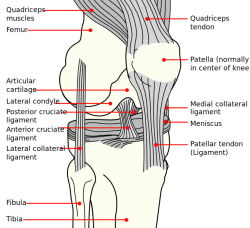
[[Image:Knee ligaments.png|250x250px|lateral collateral ligament|right|frameless]]The lateral | [[Image:Knee ligaments.png|250x250px|lateral collateral ligament|right|frameless]]The fibular or lateral collateral ligament (LCL) is the primary varus stabilizer of the knee. <ref name=":0">LaPrade, R. F., Macalena, J. A. [https://www.researchgate.net/publication/309550053_Fibular_Collateral_Ligament_and_the_Posterolateral_Corner Fibular collateral ligament and the posterolateral corner.] Insall & Scott Surgery of the Knee. 5th ed. Philadelphia, PA: Elsevier/Churchill Livingstone, 2012; 45: 592-607. | ||
</ref> It is one of 4 critical ligaments involved in stabilizing the [[Knee|knee joint.]] | |||
=== Attachments === | === Attachments === | ||
The lateral collateral ligament | The lateral collateral ligament, a cordlike band, has its origin on the lateral epicondyle of the [[femur]] and extends to the fibular head. <ref name="two">Schünke M, Schulte E, Schumacher U. Prometheus deel 1: Algemene anatomie en bewegingsapparaat. Houten: Bohn Stafleu Van Loghum, 2005.</ref><ref name="nine">Whiting WC, Zernicke RF. Biomechanics of musculoskeletal injury. 2nd ed. Human Kinetics, 2008.</ref> | ||
At the proximal level the ligament is closely related to the joint capsule, but has no direct contact with the joint capsule, it is separated by fat pad, Its distal insertion is augmented by the [[Iliotibial Band Syndrome|iliotibial tract]]. <ref name=":1">Malagelada F, Vega J, Golano P, Beynnon B, Ertem F. Knee Anatomy and Biomechanics of the Knee. In: DeLee & Drez's Orthopaedic Sports Medicine. 4th ed. Elsevier Health Sciences, 2014. | |||
</ref> | |||
== Function == | == Function == | ||
The lateral | The LCL stabilizes the lateral side of the knee joint, mainly in varus stress and posterolateral rotation of the tibia relative to the femur. LCL is one of the structures that are important as secondary stabilizers to anterior and posterior tibial translation when the cruciate ligaments are torn. <ref name=":0" /> | ||
It is primary restraint to varus rotation from 0-30° of knee flexion. As the knee goes into flexion, the LCL loses its significance and influence as a varus-stabilizing structure.<ref name=":2">Miller RH, Azar FM. Knee injuries. In: Campbell's Operative Orthopaedics. 13th ed. Philadelphia: Elsevier, 201; 2121-2297. | |||
</ref> When the knee is extended, the LCL is stretched. | |||
== Clinical relevance == | == Clinical relevance == | ||
From all knee injury’s the | From all knee injury’s the incidence of LCL is not as high as for the medial collateral ligament and it takes only 6%.<ref>Tandeter HB., Shvartzman, P. [https://www.ncbi.nlm.nih.gov/pubmed/10605994 Acute knee injuries: use of decision rules for selective radiograph ordering.] Am Fam Physician 1999; 60(9): 2599-2608.</ref> [[Lateral Collateral Ligament Injury of the Knee|Injuries to the LCL]] are commonly associated with other knee ligament injuries, thus LCL tear can be overlooked because of other concomitant injuries, and the difficulty is in the proper diagnosis. <ref>Kane PW et al. [https://www.ncbi.nlm.nih.gov/pubmed/29884567 Increased Accuracy of Varus Stress Radiographs Versus Magnetic Resonance Imaging in Diagnosing Fibular Collateral Ligament Grade III Tears.] Arthroscopy, 2018. | ||
</ref> | |||
A direct blow to the anteromedial knee is a common mechanism of injury to the LCL and posterolateral corner account, although noncontact hyperextension and noncontact varus stress injuries have also been well described.<ref name=":1" /> | |||
== Assessment == | == Assessment == | ||
=== Palpation === | === Palpation === | ||
To palpate LCL, have the patient cross his legs so that his ankle rests upon the opposite knee. When the knee is flexed to 90 degrees and the hip is abducted and externally rotated, the iliotibial tract relaxes and makes the LCL easier to isolate. The ligament lies laterally and posteriorly along the joint line. Ocassionally, the LCL is congenitally absent. <ref>Hoppenfeld S, Hutton R, Hugh T. Physical examination of the spine and extremities. 1st ed. New York: Appleton-Century-Crofts, 1976.</ref> When LCL is injured or torn, this cordlike band is not as noticeable as on the undamaged side. | |||
=== Special tests === | |||
==== <u>Adduction (Varus) stress test</u> ==== | |||
===== Purpose ===== | |||
The Varus stress test shows a lateral joint line gap. | |||
===== Performance ===== | |||
A varus stress test is performed by stabilizing the femur and palpating the lateral joint line. The other hand provides a varus stress to the ankle. The test is performed at 0 and 30 degrees, so the knee joint is in the closed packed position. While the physiotherapist with one hand is stabilizing the knee, the other hand adducts the ankle. | |||
===== Interpretation ===== | |||
If the knee joint adducts greater than normal (compare with the uninjured leg), the test is positive. This is indicative of a lateral collateral ligament tear. | |||
===== Reliability and Validity ===== | |||
* Sensitivity: 25%. Specificity: not reported. Varus stress testing was performed in 20° of flexion, and testing in extension was not done. <ref>Malanga GA, Andrus S, Nadler SF, McLean J. [https://www.archives-pmr.org/article/S0003-9993%2802%2904844-X/pdf Physical examination of the knee: a review of the original test description and scientific validity of common orthopedic tests.] Arch Phys Med Rehabil, 2003; 84(4): 592-603.</ref> | |||
* Sensitivity: 25% . The reliability of this test in extension is 68% and in 30° flexion only 56%. The test is fairly solid.<ref>Merriman L, Turner W. Assessment of the Lower Limb. 2nd ed. Churchill Livingstone, 2002.</ref> | |||
{{#ev:youtube|sg1gk6QKARw}} <ref>Physiotutors. Varus Stress Test of the Knee⎟Lateral Collateral Ligament. Available from: https://www.youtube.com/watch?v=sg1gk6QKARw </ref> | |||
==== Additional tests for detecting LCL injury with other knee ligaments: <ref name=":2" /> ==== | |||
* External Rotation-Recurvatum Test | |||
* Reverse Pivot Shift Sign of Jakob, Hassler, and Stäubli | |||
* [[Dial Test]] | |||
== Resources == | |||
Articles:<br>- Robert F. LaPrade, Spiridonov SI, Coobs BR, Ruckert PR, Griffith CJ. Fibular collateral ligament anatomical reconstructions: a prospective outcomes study. Am J Sports Med. 2010; 38: 2005-2012<br>- Benjamin R. Coobs, Robert F. LaPrade, Chad J. Griffith, Bradley J. Nelson. Biomechanical Analysis of an Isolated Fibular (Lateral) Collateral Ligament Reconstruction Using an Autogenous Semitendinosus Graft. Am J Sports Med. 2007; 35: 1521-1527<br>- DE Cooper. Tests for posterolateral instability of the knee in normal subjects. Results of examination under anesthesia. J bone Joint Surg Am. 1991; 73: 30-36<br> | Articles:<br>- Robert F. LaPrade, Spiridonov SI, Coobs BR, Ruckert PR, Griffith CJ. Fibular collateral ligament anatomical reconstructions: a prospective outcomes study. Am J Sports Med. 2010; 38: 2005-2012<br>- Benjamin R. Coobs, Robert F. LaPrade, Chad J. Griffith, Bradley J. Nelson. Biomechanical Analysis of an Isolated Fibular (Lateral) Collateral Ligament Reconstruction Using an Autogenous Semitendinosus Graft. Am J Sports Med. 2007; 35: 1521-1527<br>- DE Cooper. Tests for posterolateral instability of the knee in normal subjects. Results of examination under anesthesia. J bone Joint Surg Am. 1991; 73: 30-36<br> | ||
== References | == References == | ||
<references /> | <references /> | ||
[[Category:Knee_Anatomy]] [[Category:Vrije_Universiteit_Brussel_Project]] [[Category:Sports_Injuries]][[Category:Musculoskeletal/Orthopaedics]] [[Category:Knee]] [[Category:Assessment]] [[Category:Ligaments]] | [[Category:Knee_Anatomy]] [[Category:Vrije_Universiteit_Brussel_Project]] [[Category:Sports_Injuries]][[Category:Musculoskeletal/Orthopaedics]] [[Category:Knee]] [[Category:Assessment]] [[Category:Ligaments]] | ||
Revision as of 20:43, 9 July 2018
Original Editors - Dorien Scheirs, Joris De Pot
Top Contributors - Saimat Lachinova, Admin, Joris De Pot, Kim Jackson, Dorien Scheirs, Rachael Lowe, Leana Louw, WikiSysop, Oyemi Sillo, Kai A. Sigel, Tony Lowe, Derycker Andries, Naomi O'Reilly and George Prudden
Description[edit | edit source]
The fibular or lateral collateral ligament (LCL) is the primary varus stabilizer of the knee. [1] It is one of 4 critical ligaments involved in stabilizing the knee joint.
Attachments[edit | edit source]
The lateral collateral ligament, a cordlike band, has its origin on the lateral epicondyle of the femur and extends to the fibular head. [2][3]
At the proximal level the ligament is closely related to the joint capsule, but has no direct contact with the joint capsule, it is separated by fat pad, Its distal insertion is augmented by the iliotibial tract. [4]
Function[edit | edit source]
The LCL stabilizes the lateral side of the knee joint, mainly in varus stress and posterolateral rotation of the tibia relative to the femur. LCL is one of the structures that are important as secondary stabilizers to anterior and posterior tibial translation when the cruciate ligaments are torn. [1]
It is primary restraint to varus rotation from 0-30° of knee flexion. As the knee goes into flexion, the LCL loses its significance and influence as a varus-stabilizing structure.[5] When the knee is extended, the LCL is stretched.
Clinical relevance[edit | edit source]
From all knee injury’s the incidence of LCL is not as high as for the medial collateral ligament and it takes only 6%.[6] Injuries to the LCL are commonly associated with other knee ligament injuries, thus LCL tear can be overlooked because of other concomitant injuries, and the difficulty is in the proper diagnosis. [7]
A direct blow to the anteromedial knee is a common mechanism of injury to the LCL and posterolateral corner account, although noncontact hyperextension and noncontact varus stress injuries have also been well described.[4]
Assessment[edit | edit source]
Palpation[edit | edit source]
To palpate LCL, have the patient cross his legs so that his ankle rests upon the opposite knee. When the knee is flexed to 90 degrees and the hip is abducted and externally rotated, the iliotibial tract relaxes and makes the LCL easier to isolate. The ligament lies laterally and posteriorly along the joint line. Ocassionally, the LCL is congenitally absent. [8] When LCL is injured or torn, this cordlike band is not as noticeable as on the undamaged side.
Special tests[edit | edit source]
Adduction (Varus) stress test[edit | edit source]
Purpose[edit | edit source]
The Varus stress test shows a lateral joint line gap.
Performance[edit | edit source]
A varus stress test is performed by stabilizing the femur and palpating the lateral joint line. The other hand provides a varus stress to the ankle. The test is performed at 0 and 30 degrees, so the knee joint is in the closed packed position. While the physiotherapist with one hand is stabilizing the knee, the other hand adducts the ankle.
Interpretation[edit | edit source]
If the knee joint adducts greater than normal (compare with the uninjured leg), the test is positive. This is indicative of a lateral collateral ligament tear.
Reliability and Validity[edit | edit source]
- Sensitivity: 25%. Specificity: not reported. Varus stress testing was performed in 20° of flexion, and testing in extension was not done. [9]
- Sensitivity: 25% . The reliability of this test in extension is 68% and in 30° flexion only 56%. The test is fairly solid.[10]
Additional tests for detecting LCL injury with other knee ligaments: [5][edit | edit source]
- External Rotation-Recurvatum Test
- Reverse Pivot Shift Sign of Jakob, Hassler, and Stäubli
- Dial Test
Resources[edit | edit source]
Articles:
- Robert F. LaPrade, Spiridonov SI, Coobs BR, Ruckert PR, Griffith CJ. Fibular collateral ligament anatomical reconstructions: a prospective outcomes study. Am J Sports Med. 2010; 38: 2005-2012
- Benjamin R. Coobs, Robert F. LaPrade, Chad J. Griffith, Bradley J. Nelson. Biomechanical Analysis of an Isolated Fibular (Lateral) Collateral Ligament Reconstruction Using an Autogenous Semitendinosus Graft. Am J Sports Med. 2007; 35: 1521-1527
- DE Cooper. Tests for posterolateral instability of the knee in normal subjects. Results of examination under anesthesia. J bone Joint Surg Am. 1991; 73: 30-36
References[edit | edit source]
- ↑ 1.0 1.1 LaPrade, R. F., Macalena, J. A. Fibular collateral ligament and the posterolateral corner. Insall & Scott Surgery of the Knee. 5th ed. Philadelphia, PA: Elsevier/Churchill Livingstone, 2012; 45: 592-607.
- ↑ Schünke M, Schulte E, Schumacher U. Prometheus deel 1: Algemene anatomie en bewegingsapparaat. Houten: Bohn Stafleu Van Loghum, 2005.
- ↑ Whiting WC, Zernicke RF. Biomechanics of musculoskeletal injury. 2nd ed. Human Kinetics, 2008.
- ↑ 4.0 4.1 Malagelada F, Vega J, Golano P, Beynnon B, Ertem F. Knee Anatomy and Biomechanics of the Knee. In: DeLee & Drez's Orthopaedic Sports Medicine. 4th ed. Elsevier Health Sciences, 2014.
- ↑ 5.0 5.1 Miller RH, Azar FM. Knee injuries. In: Campbell's Operative Orthopaedics. 13th ed. Philadelphia: Elsevier, 201; 2121-2297.
- ↑ Tandeter HB., Shvartzman, P. Acute knee injuries: use of decision rules for selective radiograph ordering. Am Fam Physician 1999; 60(9): 2599-2608.
- ↑ Kane PW et al. Increased Accuracy of Varus Stress Radiographs Versus Magnetic Resonance Imaging in Diagnosing Fibular Collateral Ligament Grade III Tears. Arthroscopy, 2018.
- ↑ Hoppenfeld S, Hutton R, Hugh T. Physical examination of the spine and extremities. 1st ed. New York: Appleton-Century-Crofts, 1976.
- ↑ Malanga GA, Andrus S, Nadler SF, McLean J. Physical examination of the knee: a review of the original test description and scientific validity of common orthopedic tests. Arch Phys Med Rehabil, 2003; 84(4): 592-603.
- ↑ Merriman L, Turner W. Assessment of the Lower Limb. 2nd ed. Churchill Livingstone, 2002.
- ↑ Physiotutors. Varus Stress Test of the Knee⎟Lateral Collateral Ligament. Available from: https://www.youtube.com/watch?v=sg1gk6QKARw


