MRI Scans: Difference between revisions
Kim Jackson (talk | contribs) mNo edit summary |
No edit summary |
||
| Line 6: | Line 6: | ||
== Introduction == | == Introduction == | ||
MRI ( | MRI (an abbreviation of magnetic resonance imaging) is an imaging modality that uses non-ionising radiation to create useful diagnostic images. | ||
In simple terms, an MRI scanner consists of a large, powerful magnet in which the patient lies. A radio wave antenna is used to send signals to the body and then a radiofrequency receiver detects the emitted signals. These returning signals are converted into images by a computer attached to the scanner. Imaging of any part of the body can be obtained in any plane<ref>Radiopedia [https://radiopaedia.org/articles/mri-2?lang=gb MRI] Available from:https://radiopaedia.org/articles/mri-2?lang=gb (accessed 5.1.2021)</ref>. | |||
== Description == | == Description == | ||
MRI scan has wide range of application in medical diagnosis.<ref>Wikipedia,fckLRthe free encyclopedia http://en.wikipedia.org/wiki/Magnetic_resonance_imaging</ref> | MRI scan has wide range of application in medical diagnosis.<ref>Wikipedia,fckLRthe free encyclopedia http://en.wikipedia.org/wiki/Magnetic_resonance_imaging</ref> | ||
[[File:311.jpg|right|frameless]] | |||
'''Neuroimaging''':MRI is the investigative tool of choice for neurological cancers as it is more sensitive than CT for small tumours and offers better visualization of the posterior fossa. The contrast provided between grey and white matter makes it the optimal choice for many conditions of the central nervous system including demyelinating diseases, dementia, cerebrovascular disease, infectious diseases and epilepsy<ref>ACR–ASNR–SPR PRACTICE GUIDELINE FOR THE PERFORMANCE AND fckLRINTERPRETATION OF MAGNETIC RESONANCE IMAGING (MRI) OF THE fckLRBRAIN http://www.acr.org/~/media/ACR/Documents/PGTS/guidelines/MRI_Brain.pdf</ref> | '''Neuroimaging''': MRI is the investigative tool of choice for neurological [[Oncological Disorders|cancers]] as it is more sensitive than [[CT Scans|CT]] for small tumours and offers better visualization of the posterior fossa. The contrast provided between grey and white matter makes it the optimal choice for many conditions of the [[Introduction to Neuroanatomy|central nervous system]] including demyelinating diseases (eg [[MS Multiple Sclerosis|MS]]), [[dementia]], [[Stroke|cerebrovascular disease]], [[Infectious Disease|infectious diseases]] and [[epilepsy]]<ref>ACR–ASNR–SPR PRACTICE GUIDELINE FOR THE PERFORMANCE AND fckLRINTERPRETATION OF MAGNETIC RESONANCE IMAGING (MRI) OF THE fckLRBRAIN http://www.acr.org/~/media/ACR/Documents/PGTS/guidelines/MRI_Brain.pdf</ref> | ||
| |||
'''[[Cardiovascular System|Cardiovascular]]:''' | |||
* Cardiac MRI is complementary to other imaging techniques, such as echocardiography, cardiac CT and nuclear medicine. Its applications include assessment of [[Myocardial Infarction|myocardial ischemia]] and viability, cardiomyopathies, myocarditis, iron overload, vascular diseases and congenital heart disease<ref>"ACCF/ACR/SCCT/SCMR/ASNC/NASCI/SCAI/SIR 2006 Appropriateness Criteria for Cardiac Computed Tomography and Cardiac Magnetic Resonance Imaging". Journal of the American College of Radiology 3 (10): 751–771. 2006.</ref>. | |||
* '''Magnetic resonance angiography:''' (usually shortened to MR angiography or MRA) is an alternative to conventional angiography and CT angiography, eliminating the need for ionising radiation and iodinated contrast media, and sometimes contrast media altogether. It has evolved into several techniques with different advantages and applications: | |||
** contrast enhanced MR angiography (MRA) | |||
** non-contrast enhanced MR angiography (MRA)<ref>Radiopedia [https://radiopaedia.org/articles/mr-angiography-2?lang=gb MR angiography] Available from: https://radiopaedia.org/articles/mr-angiography-2?lang=gb<nowiki/>(accessed 5.1.2021)</ref> | |||
'''Musculoskeletal:''' Applications in the musculoskeletal system include spinal imaging, assessment of joint disease and soft tissue tumors<ref>Helms, C (2008). Musculoskeletal MRI. Saunders. ISBN 1416055347.</ref> | '''Musculoskeletal:''' Applications in the musculoskeletal system include spinal imaging, assessment of joint disease and soft tissue tumors<ref>Helms, C (2008). Musculoskeletal MRI. Saunders. ISBN 1416055347.</ref> | ||
'''Oncology:''' MRI is the investigation of choice in the preoperative staging of | '''Oncology:''' MRI is the investigation of choice in the preoperative staging of colorectal and [[Prostate Cancer|prostate cancer]], and has a role in the diagnosis, staging, and follow-up of other tumors.<ref name=":0">Giussani C, Roux FE, Ojemann J, Sganzerla EP, Pirillo D, Papagno C (2010). "Is preoperative functional magnetic resonance imaging reliable for language areas mapping in brain tumor surgery? Review of language functional magnetic resonance imaging and direct cortical stimulation correlation studies". Neurosurgery 66 (1): 113–20. doi:10.1227/01.NEU.0000360392.15450.C9. PMID 19935438.</ref> | ||
'''Liver and gastrointestinal MRI:''' Hepatobiliary MRI is used to detect and characterize lesions of the liver, pancreas and bile ducts. Focal or diffuse disorders of the liver may be evaluated using diffusion-weighted, opposed-phase imaging and dynamic contrast enhancement sequences. Extracellular contrast agents are widely used in liver MRI and newer hepatobiliary contrast agents also provide the opportunity to perform functional biliary imaging. Anatomical imaging of the bile ducts is achieved by using a heavily T2-weighted sequence in magnetic resonance cholangiopancreatography (MRCP)<ref>Frydrychowicz A, Lubner MG, Brown JJ, Merkle EM, Nagle SK, Rofsky NM, Reeder SB (2012). "Hepatobiliary MR imaging with gadolinium-based contrast agents". J Magn Reson Imaging 35 (3): 492–511. doi:10.1002/jmri.22833. PMC 3281562. PMID 22334493.</ref> | '''Liver and gastrointestinal MRI:''' Hepatobiliary MRI is used to detect and characterize lesions of the liver, pancreas and bile ducts. Focal or diffuse disorders of the liver may be evaluated using diffusion-weighted, opposed-phase imaging and dynamic contrast enhancement sequences. Extracellular contrast agents are widely used in liver MRI and newer hepatobiliary contrast agents also provide the opportunity to perform functional biliary imaging. Anatomical imaging of the bile ducts is achieved by using a heavily T2-weighted sequence in magnetic resonance cholangiopancreatography (MRCP)<ref>Frydrychowicz A, Lubner MG, Brown JJ, Merkle EM, Nagle SK, Rofsky NM, Reeder SB (2012). "Hepatobiliary MR imaging with gadolinium-based contrast agents". J Magn Reson Imaging 35 (3): 492–511. doi:10.1002/jmri.22833. PMC 3281562. PMID 22334493.</ref> | ||
| Line 43: | Line 44: | ||
==== '''Advantages of MRI''' ==== | ==== '''Advantages of MRI''' ==== | ||
* | * ability to image without the use of ionising x-rays, in contradistinction to CT scanning | ||
* | * images may be acquired in multiple planes (axial, sagittal, coronal, or oblique) without repositioning the patient. CT images have only relatively recently been able to be reconstructed in multiple planes with the same spatial resolution (i.e. isotropic voxels) | ||
* | * MRI images demonstrate superior soft-tissue contrast as compared to CT scans and plain radiographs making it the ideal examination of the brain, spine, joints, and other soft tissue body parts | ||
* | * some angiographic images can be obtained without the use of contrast material, unlike CT or conventional angiography | ||
* advanced techniques such as diffusion, spectroscopy, and perfusion allow for precise tissue characterisation rather than merely 'macroscopic' imaging | |||
* functional MRI allows visualisation of active parts of the brain during certain activities and also understanding of the underlying networks | |||
'''Disadvantages of MRI''' | '''Disadvantages of MRI''' | ||
* | * MRI scans are more expensive than CT scans | ||
* | * MRI scans take significantly longer to acquire than CT and patient comfort can be an issue, maybe exacerbated by: | ||
* | * MR image acquisition is noisy compared to CT | ||
* | * MRI scanner bores tend to be more enclosed than CT with associated claustrophobia | ||
* MR images are subject to unique artifacts that must be recognised and mitigated against (see MRI artifacts) | |||
* MRI scanning is not safe for patients with some metal implants and foreign bodies. Careful attention to safety measures is necessary to avoid serious injury to patients and staff, and this requires special MRI compatible equipment and stringent adherence to safety protocols (see MRI safety).<ref name=":0" /> | |||
=== FS – Fat Suppressed | == '''Common Abbreviations Used for MRI''' == | ||
FS – Fat Suppressed | |||
* FATSAT – Fat Saturation | * FATSAT – Fat Saturation | ||
* STIR - Short Inversion Recovery Time Imaging | * STIR - Short Inversion Recovery Time Imaging | ||
Revision as of 07:05, 5 January 2021
Original Editor - Rachael Lowe
Top Contributors - Lucinda hampton, Andeela Hafeez, Admin, Kim Jackson, Rachael Lowe, Scott Buxton, Naomi O'Reilly, Karen Wilson and Claire Knott
Introduction[edit | edit source]
MRI (an abbreviation of magnetic resonance imaging) is an imaging modality that uses non-ionising radiation to create useful diagnostic images.
In simple terms, an MRI scanner consists of a large, powerful magnet in which the patient lies. A radio wave antenna is used to send signals to the body and then a radiofrequency receiver detects the emitted signals. These returning signals are converted into images by a computer attached to the scanner. Imaging of any part of the body can be obtained in any plane[1].
Description[edit | edit source]
MRI scan has wide range of application in medical diagnosis.[2]
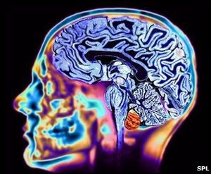
Neuroimaging: MRI is the investigative tool of choice for neurological cancers as it is more sensitive than CT for small tumours and offers better visualization of the posterior fossa. The contrast provided between grey and white matter makes it the optimal choice for many conditions of the central nervous system including demyelinating diseases (eg MS), dementia, cerebrovascular disease, infectious diseases and epilepsy[3]
- Cardiac MRI is complementary to other imaging techniques, such as echocardiography, cardiac CT and nuclear medicine. Its applications include assessment of myocardial ischemia and viability, cardiomyopathies, myocarditis, iron overload, vascular diseases and congenital heart disease[4].
- Magnetic resonance angiography: (usually shortened to MR angiography or MRA) is an alternative to conventional angiography and CT angiography, eliminating the need for ionising radiation and iodinated contrast media, and sometimes contrast media altogether. It has evolved into several techniques with different advantages and applications:
- contrast enhanced MR angiography (MRA)
- non-contrast enhanced MR angiography (MRA)[5]
Musculoskeletal: Applications in the musculoskeletal system include spinal imaging, assessment of joint disease and soft tissue tumors[6]
Oncology: MRI is the investigation of choice in the preoperative staging of colorectal and prostate cancer, and has a role in the diagnosis, staging, and follow-up of other tumors.[7]
Liver and gastrointestinal MRI: Hepatobiliary MRI is used to detect and characterize lesions of the liver, pancreas and bile ducts. Focal or diffuse disorders of the liver may be evaluated using diffusion-weighted, opposed-phase imaging and dynamic contrast enhancement sequences. Extracellular contrast agents are widely used in liver MRI and newer hepatobiliary contrast agents also provide the opportunity to perform functional biliary imaging. Anatomical imaging of the bile ducts is achieved by using a heavily T2-weighted sequence in magnetic resonance cholangiopancreatography (MRCP)[8]
Working[edit | edit source]
MRI uses three electromagnetic fields: a very strong (on the order of units of teslas) static magnetic field to polarize the hydrogen nuclei, called the static field; a weaker time-varying (on the order of 1 kHz) field(s) for spatial encoding, called the gradient field(s); and a weak radio-frequency (RF) field for manipulation of the hydrogen nuclei to produce measurable signals, collected through an RF antenna.
Procedure[edit | edit source]
During an MRI scan, you lie on a flat bed that is moved into the scanner. Depending on the part of your body being scanned, you will be moved into the scanner either head first or feet first.
The MRI scanner is operated by a radiographer, who is trained in carrying out X-rays and similar procedures. They control the scanner using a computer, which is in a different room to keep it away from the magnetic field generated by the scanner.
You will be able to talk to the radiographer through an intercom and they will be able to see you on a television monitor throughout the scan.
At certain times during the scan, the scanner will make loud tapping noises. This is the electric current in the scanner coils being turned on and off. You will be given earplugs or headphones to wear.
It is very important that you keep as still as possible during your MRI scan. The scan will last between 15 and 90 minutes, depending on the size of the area being scanned and how many images are taken[9]
Advantages and Disadvantages of MRI[edit | edit source]
Advantages of MRI[edit | edit source]
- ability to image without the use of ionising x-rays, in contradistinction to CT scanning
- images may be acquired in multiple planes (axial, sagittal, coronal, or oblique) without repositioning the patient. CT images have only relatively recently been able to be reconstructed in multiple planes with the same spatial resolution (i.e. isotropic voxels)
- MRI images demonstrate superior soft-tissue contrast as compared to CT scans and plain radiographs making it the ideal examination of the brain, spine, joints, and other soft tissue body parts
- some angiographic images can be obtained without the use of contrast material, unlike CT or conventional angiography
- advanced techniques such as diffusion, spectroscopy, and perfusion allow for precise tissue characterisation rather than merely 'macroscopic' imaging
- functional MRI allows visualisation of active parts of the brain during certain activities and also understanding of the underlying networks
Disadvantages of MRI
- MRI scans are more expensive than CT scans
- MRI scans take significantly longer to acquire than CT and patient comfort can be an issue, maybe exacerbated by:
- MR image acquisition is noisy compared to CT
- MRI scanner bores tend to be more enclosed than CT with associated claustrophobia
- MR images are subject to unique artifacts that must be recognised and mitigated against (see MRI artifacts)
- MRI scanning is not safe for patients with some metal implants and foreign bodies. Careful attention to safety measures is necessary to avoid serious injury to patients and staff, and this requires special MRI compatible equipment and stringent adherence to safety protocols (see MRI safety).[7]
Common Abbreviations Used for MRI[edit | edit source]
FS – Fat Suppressed
- FATSAT – Fat Saturation
- STIR - Short Inversion Recovery Time Imaging
- FSE – Fast Spin Echo
- Gad – Gadolinium
Hybrid MRI Sequences occurs with manipulation to the type and frequency of radio frequency and cause echoes. A gradient echo adds sensitivity or iron-complexes such as articular cartilage defects and haemorrhaging in muscle, but conversely decreases resolution on metal hardware (such as pins or screws) from a surgery. Spin echo adds the benefit of increased tissue contrast and better visualisation of meniscal tears. Stimulated echo reduce interference of signal and therefore may be used to look at specific molecular movements within tissue.[5]
T1 Weighted MRI
- Demonstrates good anatomic structures
- Fat and meniscal tears appear bright white
- Water, CSF, muscle, tendons, and ligaments appears light to dark grey
- Air and cortical bone appears dark
T2 Weighted MRI
- Demonstrates contrast between normal and abnormal (can identify abnormal lesions of fluid)
- Water and CSF appears bright white
- Fat, muscle, tendons, ligaments and cartilage appear light to dark gray
- Air and cortical bone appears dark (unless fluid is in the lung)
Proton density images
- An image simply of the density of protons
- A more dense area of protons will appear white (cortical bone, bone marrow)
- A less dense region will appear darker (fluids, soft tissues)
Contraindications for MRI
- Pacemakers, aneurysm clips, cochlear implants, and orbital foreign bodies
- Projectiles in the room (includes oxygen tanks, IV poles, stethoscopes, hair pins, etc)
Difference between MRI and CT[edit | edit source]
Like CT, MRI traditionally creates a two dimensional image of a thin "slice" of the body and is therefore considered a tomographic imaging technique. Modern MRI instruments are capable of producing images in the form of 3D blocks, which may be considered a generalisation of the single-slice, tomographic, concept. Unlike CT, MRI scans do not use X-rays so the possible concerns associated with X-ray pictures and CT scans (which use X-rays) are not associated with MRI scans.[10] For example, because MRI has only been in use since the early 1980s, there are no known long-term effects of exposure to strong static fields (this is the subject of some debate; see 'Safety' in MRI) and therefore there is no limit to the number of scans to which an individual can be subjected, in contrast with X-ray and CT. However, there are well-identified health risks associated with tissue heating from exposure to the RF field and the presence of implanted devices in the body, such as pacemakers. These risks are strictly controlled as part of the design of the instrument and the scanning protocols used.
Because CT and MRI are sensitive to different tissue properties, the appearance of the images obtained with the two techniques differs markedly. In CT, X-rays must be blocked by some form of dense tissue to create an image, so the image quality when looking at soft tissues will be poor. In MRI, while any nucleus with a net nuclear spin can be used, the proton of the hydrogen atom remains the most widely used, especially in the clinical setting, because it is so ubiquitous and returns a large signal. This nucleus, present in water molecules, allows the excellent soft-tissue contrast achievable.
Reference[edit | edit source]
- ↑ Radiopedia MRI Available from:https://radiopaedia.org/articles/mri-2?lang=gb (accessed 5.1.2021)
- ↑ Wikipedia,fckLRthe free encyclopedia http://en.wikipedia.org/wiki/Magnetic_resonance_imaging
- ↑ ACR–ASNR–SPR PRACTICE GUIDELINE FOR THE PERFORMANCE AND fckLRINTERPRETATION OF MAGNETIC RESONANCE IMAGING (MRI) OF THE fckLRBRAIN http://www.acr.org/~/media/ACR/Documents/PGTS/guidelines/MRI_Brain.pdf
- ↑ "ACCF/ACR/SCCT/SCMR/ASNC/NASCI/SCAI/SIR 2006 Appropriateness Criteria for Cardiac Computed Tomography and Cardiac Magnetic Resonance Imaging". Journal of the American College of Radiology 3 (10): 751–771. 2006.
- ↑ Radiopedia MR angiography Available from: https://radiopaedia.org/articles/mr-angiography-2?lang=gb(accessed 5.1.2021)
- ↑ Helms, C (2008). Musculoskeletal MRI. Saunders. ISBN 1416055347.
- ↑ 7.0 7.1 Giussani C, Roux FE, Ojemann J, Sganzerla EP, Pirillo D, Papagno C (2010). "Is preoperative functional magnetic resonance imaging reliable for language areas mapping in brain tumor surgery? Review of language functional magnetic resonance imaging and direct cortical stimulation correlation studies". Neurosurgery 66 (1): 113–20. doi:10.1227/01.NEU.0000360392.15450.C9. PMID 19935438.
- ↑ Frydrychowicz A, Lubner MG, Brown JJ, Merkle EM, Nagle SK, Rofsky NM, Reeder SB (2012). "Hepatobiliary MR imaging with gadolinium-based contrast agents". J Magn Reson Imaging 35 (3): 492–511. doi:10.1002/jmri.22833. PMC 3281562. PMID 22334493.
- ↑ Go to NHS Choices homepageYour health, your choices http://www.nhs.uk/conditions/MRI-scan/Pages/Introduction.aspx
- ↑ patient co uk trusted medical information and support http://www.patient.co.uk/health/mri-scan


