Median Nerve: Difference between revisions
No edit summary |
No edit summary |
||
| Line 12: | Line 12: | ||
In the forearm it passes between the two heads of the Pronator teres and crosses the ulnar artery, but is separated from this vessel by the deep head of the Pronator teres. It descends beneath the Flexor digitorum sublimis, lying on the Flexor digitorum profundus, to within 5 cm. of the transverse carpal ligament; here it becomes more superficial, and is situated between the tendons of the Flexor digitorum sublimis and Flexor carpi radialis. | In the forearm it passes between the two heads of the Pronator teres and crosses the ulnar artery, but is separated from this vessel by the deep head of the Pronator teres. It descends beneath the Flexor digitorum sublimis, lying on the Flexor digitorum profundus, to within 5 cm. of the transverse carpal ligament; here it becomes more superficial, and is situated between the tendons of the Flexor digitorum sublimis and Flexor carpi radialis. | ||
In this situation it lies behind, and rather to the radial side of, the tendon of the Palmaris longus, and is covered by the skin and [[Fascia|fascia]]. It then passes behind the transverse carpal ligament into the palm of the hand. In its course through the forearm it is accompanied by the median artery, a branch of the volar interroseous artery. <ref name="Grey">Gray, Henry. Anatomy of the human body. Philadelphia and New York: Lea & | In this situation it lies behind, and rather to the radial side of, the tendon of the Palmaris longus, and is covered by the skin and [[Fascia|fascia]]. It then passes behind the transverse carpal ligament into the palm of the hand. In its course through the forearm it is accompanied by the median artery, a branch of the volar interroseous artery. <ref name="Grey">Gray, Henry. Anatomy of the human body. Philadelphia and New York: Lea &amp; Febiger, 1825-1861</ref> <br> | ||
=== Root === | === Root === | ||
| Line 32: | Line 32: | ||
*palmar | *palmar | ||
'''Variation''' | |||
*martin gruber anastomosis; | *martin gruber anastomosis; | ||
| Line 43: | Line 43: | ||
=== Motor === | === Motor === | ||
'''Muscular branch''' | |||
*all the superficial muscles on the front of the forearm except the flexor carpi ulnaris | *all the superficial muscles on the front of the forearm except the flexor carpi ulnaris | ||
Volar interosseous branch | '''Volar interosseous branch''' | ||
*deep muscles on the front of the forearm, except the ulnar half of the Flexor digitorum profundus | *deep muscles on the front of the forearm, except the ulnar half of the Flexor digitorum profundus | ||
| Line 53: | Line 53: | ||
=== <br> Sensory === | === <br> Sensory === | ||
'''Palmar branch''' | |||
It pierces the volar carpal ligament, and divides into: | It pierces the volar carpal ligament, and divides into: | ||
| Line 59: | Line 59: | ||
*lateral branch | *lateral branch | ||
skin over the ball of the thumb; | - skin over the ball of the thumb; | ||
communicates with the volar branch of the lateral antibrachial cutaneous nerve. | - communicates with the volar branch of the lateral antibrachial cutaneous nerve. | ||
*medial branch | *medial branch | ||
skin of the palm; | - skin of the palm; | ||
communicates with the palmar cutaneous branch of the ulnar. <ref name="Grey" /> | - communicates with the palmar cutaneous branch of the ulnar. <ref name="Grey" /> | ||
== Clinical relevance == | == Clinical relevance == | ||
| Line 79: | Line 79: | ||
=== Neuro dynamics === | === Neuro dynamics === | ||
Extending the elbow and wrist, two key components of the upper limb tension test, puts the median nerve under tension. Rotating the head and neck to the opposite side puts the nerve under increasing stretch. If the entrapment is in the inter scalene triangle then raising the arm above the head usually increases the response. The purpose is to test for C5, C6, C7 nerve roots and median nerve as the source of the patient’s painful shoulder and arm. | Extending the elbow and wrist, two key components of the upper limb tension test, puts the median nerve under tension. Rotating the head and neck to the opposite side puts the nerve under increasing stretch. If the entrapment is in the inter scalene triangle then raising the arm above the head usually increases the response. The purpose is to test for C5, C6, C7 nerve roots and median nerve as the source of the patient’s painful shoulder and arm.<ref>Special Tests. Orthopedic Testing Procedure http://special-tests.com/shoulder-tests/ultt/ (accessed 7 april 2017)</ref> | ||
'''Upper Limb Tension Test 1 (ULTT1, Median nerve bias)''' | |||
#Shoulder girdle depression | #Shoulder girdle depression | ||
| Line 90: | Line 90: | ||
#Elbow extension | #Elbow extension | ||
'''Upper Limb Tension Test 2A (ULTT2A, Median nerve bias)''' | |||
#Shoulder girdle depression | #Shoulder girdle depression | ||
#Elbow extension | #Elbow extension | ||
#Lateral rotation of the whole arm | #Lateral rotation of the whole arm | ||
#Wrist, finger and thumb extension | #Wrist, finger and thumb extension<br> | ||
<br> | |||
== Treatment<br> == | == Treatment<br> == | ||
| Line 105: | Line 103: | ||
{| width="100%" cellspacing="1" cellpadding="1" | {| width="100%" cellspacing="1" cellpadding="1" | ||
|- | |- | ||
| {{#ev:youtube|8iYxrZKAZU|300}}<ref>Kenhub. Median Nerve - Distribution, Innervation & Anatomy - Human Anatomy. Kenhub Available from: https://www.youtube.com/watch?v=-8iYxrZKAZU [last accessed 4/7/17]</ref> | | {{#ev:youtube|8iYxrZKAZU|300}}<ref>Kenhub. Median Nerve - Distribution, Innervation &amp; Anatomy - Human Anatomy. Kenhub Available from: https://www.youtube.com/watch?v=-8iYxrZKAZU [last accessed 4/7/17]</ref> | ||
| {{#ev:youtube|PvXaBrZOeIw|300}}<ref> ehowhealt. Carpal Tunnel: Median Nerve Stretches for Relieving Carpal Tunnel Syndrome Pain. Available from: https://www.youtube.com/watch?v=PvXaBrZOeIw [last accessed 4/7/17 | | {{#ev:youtube|PvXaBrZOeIw|300}}<ref> ehowhealt. Carpal Tunnel: Median Nerve Stretches for Relieving Carpal Tunnel Syndrome Pain. Available from: https://www.youtube.com/watch?v=PvXaBrZOeIw [last accessed 4/7/17]</ref> | ||
|} | |} | ||
== See also == | |||
*[[Carpal Tunnel Syndrome|Carpal Tunnel Syndrome]] | |||
*[[Brachial plexus injury|Brachial plexus injury]] | |||
*[[Neurodynamic Assessment|Neurodynamic Assessment]] | |||
== Recent Related Research (from Pubmed) == | |||
<div class="researchbox"><rss>https://eutils.ncbi.nlm.nih.gov/entrez/eutils/erss.cgi?rss_guid=1Hw5AZzsGCaE4m-n6jsgck81U3WMdTZK8sEYE2AcFLPqfH9EJG|charset=UTF-8|short|max=10</rss></div> | |||
== References == | |||
<references /> | |||
[[Category:Anatomy]] [[Category:Nerves]] | |||
Revision as of 22:07, 7 April 2017
Original Editor - Daniele Barilla
Top Contributors - Daniele Barilla, Shreya Trivedi, Naomi O'Reilly, Kim Jackson, Chrysolite Jyothi Kommu, Blessed Denzel Vhudzijena, Evan Thomas, Joao Costa, Ahmed M Diab, David Olukayode, 127.0.0.1, WikiSysop and Manisha Shrestha
Description[edit | edit source]
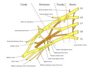
The Median Nerve extends along the middle of the arm and forearm to the hand. It arises by two roots, one from the lateral and one from the medial cord of the brachial plexus; these embrace the lower part of the axillary artery, uniting either in front of or lateral to that vessel. Its fibers are derived from the sixth, seventh, and eighth cervical and first thoracic nerves.
As it descends through the arm, it lies at first lateral to the brachial artery; about the level of the insertion of the Coracobrachialis it crosses the artery, usually in front of, but occasionally behind it, and lies on its medial side at the bend of the elbow, where it is situated behind the lacertus fibrosus (bicipital fascia), and is separated from the elbow-joint by the brachialis.
In the forearm it passes between the two heads of the Pronator teres and crosses the ulnar artery, but is separated from this vessel by the deep head of the Pronator teres. It descends beneath the Flexor digitorum sublimis, lying on the Flexor digitorum profundus, to within 5 cm. of the transverse carpal ligament; here it becomes more superficial, and is situated between the tendons of the Flexor digitorum sublimis and Flexor carpi radialis.
In this situation it lies behind, and rather to the radial side of, the tendon of the Palmaris longus, and is covered by the skin and fascia. It then passes behind the transverse carpal ligament into the palm of the hand. In its course through the forearm it is accompanied by the median artery, a branch of the volar interroseous artery. [1]
Root[edit | edit source]
C5-C6-C7-C8-T1
From[edit | edit source]
lateral and medial cords of the brachial plexus
Branches[edit | edit source]
With the exception of the nerve to the Pronator teres, which sometimes arises above the elbow-joint, the median nerve gives off no branches in the arm. As it passes in front of the elbow, it supplies one or two twigs to the joint.
In the forearm its branches are:
- muscular
- volar interosseous
- palmar
Variation
- martin gruber anastomosis;
- bifid (high division) of median nerve: associated w/ a median artery [2]
Function[edit | edit source]
Motor[edit | edit source]
Muscular branch
- all the superficial muscles on the front of the forearm except the flexor carpi ulnaris
Volar interosseous branch
- deep muscles on the front of the forearm, except the ulnar half of the Flexor digitorum profundus
Sensory[edit | edit source]
Palmar branch
It pierces the volar carpal ligament, and divides into:
- lateral branch
- skin over the ball of the thumb;
- communicates with the volar branch of the lateral antibrachial cutaneous nerve.
- medial branch
- skin of the palm;
- communicates with the palmar cutaneous branch of the ulnar. [1]
Clinical relevance[edit | edit source]
Assessment
[edit | edit source]
Neuro exams[edit | edit source]
Signs of a median nerve lesion include weak pronation of the forearm, weak flexion & radial deviation of wrist, with thenar atrophy & inability to oppose or flex the thumb;
- sensory distribution includes thumb, radial 2 1/2 fingers, and corresponding portion of palm.
- w/ intact nerve, thumb can be pronated, lining up nails at or near 180 deg;
- w/ median nerve palsy, thumb can't be pronated & nail is < 100 deg [2]
Neuro dynamics[edit | edit source]
Extending the elbow and wrist, two key components of the upper limb tension test, puts the median nerve under tension. Rotating the head and neck to the opposite side puts the nerve under increasing stretch. If the entrapment is in the inter scalene triangle then raising the arm above the head usually increases the response. The purpose is to test for C5, C6, C7 nerve roots and median nerve as the source of the patient’s painful shoulder and arm.[3]
Upper Limb Tension Test 1 (ULTT1, Median nerve bias)
- Shoulder girdle depression
- Shoulder joint abduction
- Forearm supination
- Wrist and finger extension
- Shoulder joint laterally rotated
- Elbow extension
Upper Limb Tension Test 2A (ULTT2A, Median nerve bias)
- Shoulder girdle depression
- Elbow extension
- Lateral rotation of the whole arm
- Wrist, finger and thumb extension
Treatment
[edit | edit source]
Resources[edit | edit source]
| [4] | [5] |
See also[edit | edit source]
Recent Related Research (from Pubmed)[edit | edit source]
References[edit | edit source]
- ↑ 1.0 1.1 Gray, Henry. Anatomy of the human body. Philadelphia and New York: Lea & Febiger, 1825-1861
- ↑ 2.0 2.1 Wheeless' Textbooks of Orthopaedics, Medial Nerve http://www.wheelessonline.com/ortho/Median_nerve (accessed 7 april 2017)
- ↑ Special Tests. Orthopedic Testing Procedure http://special-tests.com/shoulder-tests/ultt/ (accessed 7 april 2017)
- ↑ Kenhub. Median Nerve - Distribution, Innervation & Anatomy - Human Anatomy. Kenhub Available from: https://www.youtube.com/watch?v=-8iYxrZKAZU [last accessed 4/7/17]
- ↑ ehowhealt. Carpal Tunnel: Median Nerve Stretches for Relieving Carpal Tunnel Syndrome Pain. Available from: https://www.youtube.com/watch?v=PvXaBrZOeIw [last accessed 4/7/17]


