Median Nerve
This article is currently under review and may not be up to date. Please come back soon to see the finished work! (8/12/2021)
Original Editor - Wendy Walker and Daniele Barilla
Top Contributors - Daniele Barilla, Shreya Trivedi, Naomi O'Reilly, Kim Jackson, Chrysolite Jyothi Kommu, Blessed Denzel Vhudzijena, Evan Thomas, Joao Costa, Ahmed M Diab, David Olukayode, 127.0.0.1, WikiSysop and Manisha Shrestha
Anatomy[edit | edit source]
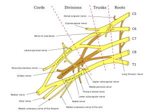
The Median Nerve begins in axillary region , root of median nerves are situated in anterior rami of C5-T1. Median and lateral cords of brachial plexus are merged and extended as median nerve. [1] Uniting either in front of or lateral to that vessel. Its fibers are derived from the sixth, seventh, and eighth cervical and first thoracic nerves.
As it descends through the arm, it lies at first lateral to the brachial artery; about the level of the insertion of the Coracobrachialis it crosses the artery, usually in front of, but occasionally behind it, and lies on its medial side at the bend of the elbow, where it is situated behind the lacertus fibrosus (bicipital fascia) and is separated from the elbow-joint by the brachialis.
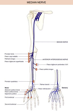
In the forearm, it passes between the two heads of the Pronator teres and crosses the ulnar artery, but is separated from this vessel by the deep head of the Pronator Teres. It descends beneath the Flexor digitorum sublimis, lying on the Flexor digitorum profundus, to within 5 cm. of the transverse carpal ligament; here it becomes more superficial and is situated between the tendons of the Flexor digitorum sublimis and Flexor Carpi Radialis.
In this situation, it lies behind, and rather to the radial side of the tendon of the Palmaris Longus, and is covered by the skin and fascia. It then passes behind the transverse carpal ligament into the palm of the hand. In its course through the forearm, it is accompanied by the median artery, a branch of the volar interosseous artery. [2]
Video[edit | edit source]
Median Nerve 3D Anatomy Tutorial
Root[edit | edit source]
C5-C6-C7-C8-T1
From[edit | edit source]
C5 to C7 roots from lateral cord of brachial plexus
C8 and T1 roots from medial cord of brachial plexus
Branches[edit | edit source]
With the exception of the nerve to the Pronator teres, which sometimes arises above the elbow-joint, the median nerve gives off no branches in the arm. As it passes in front of the elbow, it supplies one or two twigs to the joint.
| Branches of median nerve in forearm | Muscle supplied by |
|---|---|
| Muscular Branch | All the superficial muscles on the front of the forearm except the Flexor Carpi Ulnaris
|
| Anterior Interosseus Nerve (AIN) | AIN which supplies
This branch runs on the interosseous membrane with the anterior interosseous artery which supplies
It terminates by giving articular branches to the distal radioulnar, radiocarpal and carpal joints. |
| Branches of median nerve in Hand | Muscle supplied by |
| The cutaneous nerve | which supplies the proximal aspect of the palm. This branch does not enter the carpal tunnel and is hence spared in carpal tunnel syndrome. |
| Palmar digital nerves | The two common palmar digital nerves
|
| Recurrent branch
(‘million dollar nerve’ ) |
Muscles of the thenar eminence (flexor pollicis brevis, abductor pollicis brevis, opponens pollicis). |
Function[edit | edit source]
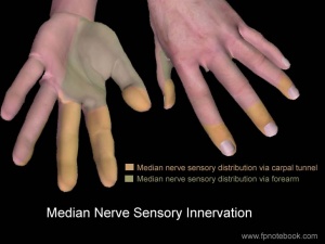
The main two function of median nerve are Motor and Sensory, the muscles supplied by median nerve are mainly flexor aspect of forearm, hand, and thumb. Sensory innervation to the dorsal aspect of the distal first two digits of the hand is supplied by median nerve. The motor function is described in table above
Sensory Function[edit | edit source]
- The skin of palamr and dorsal aspect of lateral three to three and half palm
- The skin of palmar and dorsal aspect of thumb
- The skin of adjacent sides of respective digits.
- The Skin of center of the palm
Clinical relevance[edit | edit source]
There are three entrapment syndromes involving the medial nerve or its branches:
- Carpal Tunnel Syndrome
- Anterior Interosseous Syndrome
- Pronator Teres Syndrome
Carpal Tunnel Syndrome[edit | edit source]
Is a common condition that causes a tingling sensation, numbness and sometimes pain in the hand and fingers. These sensations usually develop gradually and start off being worse during the night. They tend to affect the thumb, index finger, and middle finger.
Other symptoms of carpal tunnel syndrome include:
Anterior Interosseous Syndrome[edit | edit source]
AINS is a pure motor neuropathy, as the anterior interosseous nerve contains no sensory fibers; dull forearm pain is however sometimes mentioned by patients.
Typically, patients fail to make an “O.K.”-sign, as flexion of the interphalangeal joint of the thumb and the distal interphalangeal joint of the index finger, is impaired.
Another sensitive test is the pinch test: a patient with AINS will also not be able to pinch a sheet of paper between his thumb and index finger, instead of clamping the sheet between his extended thumb and index fingers, akin to a tong rather than a clamp. The weakness of the pronator quadratus muscle manifests itself in pronation weakness with a flexed elbow.
AINS can be confounded by the Martin-Gruber anastomosis, present in up to 25% of the population: in these cases, the anterior interosseous nerve gives off branches to the ulnar nerve, creating atypical motor innervation patterns of the forearm and hand and thus effacing the typical clinical symptoms.
Pronator Teres Syndrome[edit | edit source]
Due to the position of this muscle that sits over your median nerve, the pressure on the nerve can elicit pain while hindering your forearm movement. It occurs when your pronator teres muscle becomes tight or overworked, compressing the median nerve. Hammering, repeatedly using a screwdriver, cleaning fish can lead to overuse of the pronator teres. Symptoms include pain and reduced mobility. Carpal Tunnel Syndrome symptoms are often confused with PTS, however in this case symptoms worsen with elbow movements. Pronator Teres Syndrome test can be used for assessment of the syndrome.
Pathology/Injury[edit | edit source]
Carpal Tunnel Syndrome is a condition which occurs due to pressure on the median nerve as it passes through the carpal tunnel
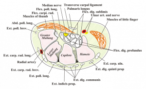
Because of anatomical structures arrangement at the writs joint the nerve become more prone to the injury at that level. The Carpal tunnel is formed by two layers, deep carpal arch and superficial flexor retinaculum. 9 tendons and median nerve pass through this tunnel. Wrist joint is most function joint and thus is moves numerous times while doing many activity which makes median nerve at this level more vulnerable to injury.[6]
Injury at the elbow[edit | edit source]
Most common injury = Supracondylar fracture of the humerus.[7]
This results in the flexors and pronators in the forearm being paralyzed, with the exception of the flexor carpi ulnaris and medial half of flexor digitorum profundus.
The forearm constantly supinated, and flexion is weak (often accompanied by adduction, because of the pull of the flexor carpi ulnaris).
Flexion at the thumb is also prevented, as both the longus and brevis muscles are paralysed.
Sensory functions: Gives rise to the palmar cutaneous branch, which innervates the lateral part of the palm, and the digital cutaneous branch, which innervates the lateral three and a half fingers on the anterior (palmar) surface of the hand.
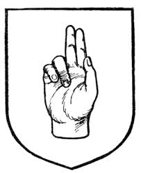
The lateral two lumbrical muscles are paralyzed, and the patient will not be able to flex at the MCP joints or extend at IP joints of the index and middle fingers. So on trying to flex the fingers and thumb to make a fist, the 1st 3 digits remain in extension, and the posture is known as the "hand of benediction".
NB In lesions of the Ulnar Nerve the same posture occurs with the hand at rest, due to paralysis of the medial lumbricals. In these cases, it is known as the "ulnar claw".
Injury at the wrist[edit | edit source]
Most common injury = Lacerations just proximal to the flexor retinaculum.[7]
Motor functions: Thenar muscles paralysed, as are the lateral two lumbricals. This affects the opposition of the thumb and flexion of the index and middle fingers.
Sensory functions: Same as an injury at the elbow.
Characteristic signs: Same as an injury at the elbow, i.e. hand of benediction on attempting to make a fist.
Clinical examination[edit | edit source]
Neuro exams[edit | edit source]
Signs of a median nerve lesion include weak pronation of the forearm, weak flexion and radial deviation of wrist, with thenar atrophy and inability to oppose or flex the thumb;
- sensory distribution includes thumb, radial 2 1/2 fingers, and the corresponding portion of palm.
- w/ intact nerve, thumb e thcan be pronated, lining up nails at or near 180 deg;
- w/ median nerve palsy, thumb can't be pronated & nail is < 100 deg
Neurodynamics[edit | edit source]
Extending the elbow and wrist, two key components of the upper limb tension test, puts the median nerve under tension. Rotating the head and neck to the opposite side puts the nerve under increasing stretch. If the entrapment is in the inter scalene triangle then raising the arm above the head usually increases the response. The purpose is to test for C5, C6, C7 nerve roots and median nerve as the source of the patient’s painful shoulder and arm.[8]
Upper Limb Tension Test 1 (ULTT1, Median nerve bias)
- Shoulder girdle depression
- Shoulder joint abduction
- Forearm supination
- Wrist and finger extension
- Shoulder joint laterally rotated
- Elbow extension
Upper Limb Tension Test 2A (ULTT2A, Median nerve bias)
- Shoulder girdle depression
- Elbow extension
- Lateral rotation of the whole arm
- Wrist, finger and thumb extension
Treatment[edit | edit source]
- Manual Therapy
- Soft Tissue Mobilization
- Neurodynamic techniques
- Functional massage
- Carpal bone mobilization techniques
- Electrophysical therapy (EM)
- Ultrasound therapy
- Laser therapy
Both therapies had a positive effect on nerve conduction, pain reduction, functional status, and subjective symptoms in individuals with CTS. However, the results regarding pain reduction, subjective symptoms, and functional status were better in the MT group.[9]
How to examine the functions of median nerve.
See also[edit | edit source]
References[edit | edit source]
- ↑ Abdalbary, S., Abdel-Wahed, M., Amr, S., Mahmoud, M., El-Shaarawy, E., Salaheldin, S. and Fares, A., 2021. The Myth of Median Nerve in Forearm and Its Role in Double Crush Syndrome: A Cadaveric Study.
- ↑ Gray, Henry. Anatomy of the human body. Philadelphia and New York: Lea & Febiger, 1825-1861
- ↑ nhs.uk. 2021. Carpal tunnel syndrome. [online] Available at: <https://www.nhs.uk/conditions/carpal-tunnel-syndrome/>
- ↑ Orthoinfo.aaos.org. 2021. [online] Available at: <https://orthoinfo.aaos.org/globalassets/pdfs/carpel-tunnel-syndrome.pdf> [Accessed 30 November 2021].
- ↑ Ncbi.nlm.nih.gov. 2021. [online] Available at: <https://www.ncbi.nlm.nih.gov/pmc/articles/PMC4727604/pdf/Neurosciences-20-4.pdf> [Accessed 30 November 2021].
- ↑ Abdalbary, S., Abdel-Wahed, M., Amr, S., Mahmoud, M., El-Shaarawy, E., Salaheldin, S. and Fares, A., 2021. The Myth of Median Nerve in Forearm and Its Role in Double Crush Syndrome: A Cadaveric Study.
- ↑ 7.0 7.1 Dydyk AM, Negrete G, Cascella M. Median Nerve Injury. [Updated 2021 Aug 25]. In: StatPearls [Internet]. Treasure Island (FL): StatPearls Publishing; 2021 Jan-. Available from: https://www.ncbi.nlm.nih.gov/books/NBK553109/
- ↑ Special Tests. 2021. Upper Limb Tension Test 1 - Special Tests. [online] Available at: <https://special-tests.com/shoulder-tests/ultt/> [Accessed 30 November 2021].
- ↑ Wolny T, Saulicz E, Linek P, Shacklock M, Mysliwiec A. Efficacy of Manual Therapy Including Neurodynamic Techniques for the Treatment of Carpal Tunnel Syndrome: A Randomized Controlled Trial. J Manipulative Physiol Ther. 2017.
