Occipital Bone: Difference between revisions
Mande Jooste (talk | contribs) No edit summary |
Mande Jooste (talk | contribs) No edit summary |
||
| Line 8: | Line 8: | ||
[[File:Fullsizeoutput_c1.jpeg|240x240px]] | [[File:Fullsizeoutput_c1.jpeg|240x240px]] | ||
[[File:Occipital_bone.png|center|frameless|480x480px]] | [[File:Occipital_bone.png|center|frameless|480x480px]] | ||
=== Articulations === | === Articulations === | ||
Revision as of 12:58, 2 April 2019
Original Editor- Hannah Hassel Top Contributors - Mande Jooste, Hannah Hassell, Kim Jackson, Lucinda hampton and Tony Lowe
Description[edit | edit source]
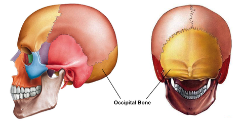
The Occipital bone is a trapezoidal-shaped bone forming the base of the skull. It is situated at the the lower and back part of the cranium. The large oval opening in the bone is called the foramen magnum through which the spinal cord exits the cranial vault.
Articulations[edit | edit source]
The occipital bone articulate with six bones:
- Two temporal bones
- Two parietal bones
- Sphenoid bone
- Atlas
Muscle attachments[edit | edit source]
Superior curved line: Occipito frontalis; Trapezius; Sternocleidomastoid
Space between the curved lines: Complexus; Splenius capitis; Obliquus superior
Inferior curved line and space between it and the foramen magnum: Rectus posticus major and minor
Transverse process: Rectus lateralis
Basilar process: Rectus antics major and minor; Superior constrictor of pharynx[1]
Clinical relevance[edit | edit source]
Basilar skull fracture
Resources[edit | edit source]
[edit | edit source]
Occipital neuralgia[2]


