Osgood-Schlatter Disease
Original Editor - Casey Kirkes, Geoffrey De Vos as part of the Vrije Universiteit Brussel Evidence-based Practice Project
Top Contributors - Geoffrey De Vos, Admin, Keta Parikh, Sam Verhelpen, Nicolas D'Hondt, Kim Jackson, Michelle Lee, Scott Buxton, Laurens Dereymaeker, Jetse De Proft, Rachael Lowe, Wanda van Niekerk, Nicolas Everaerts, Casey Kirkes, Nikhil Benhur Abburi, Rucha Gadgil, Lucinda hampton, Jess Bell, Andeela Hafeez, Hanne Velghe, Uchechukwu Chukwuemeka, Vidya Acharya, Adam Vallely Farrell, 127.0.0.1, Tony Lowe, Claire Knott, Naomi O'Reilly and Rishika BabburuDefinition/Description[edit | edit source]
Osgood-Schlatter's disease also known as osteochondrosis or traction apophysitis at the level of the tibial tubercle due to repetitive strain on the growth plate of the tibial tuberosity. The repetitive strain is from the strong pull of the quadriceps muscle(extensor mechanism) produced during sporting activities.
Sports like:
- Basketball
- Volleyball
- Sprinters
- Gymnastic
- Football
ETIOLOGY:
The Patellar tendon inserts at tibial tubercle. Repeated traction and microtrauma at the insertion causes microvascular tear, inflammation and fracture. Osgood-Schlatter disease, is an overuse injury characterized by ossification of bone along the growth plate, the tibial tubercle. Due to excessive growth, the ability of the muscle-tendon unit to stretch sufficiently and maintain flexibility is hampered, and there is increased tension across the tibial tubercle apophysis. In severe cases, repetitive strain may lead to softening and partial avulsion of the tibial tubercle apophysis resulting into osteochondritis.[1]
EPIDEMIOLOGY
- High prevalence in immature adolescent
- Most common in males, that engage in running, jumping and sprinting activities.
- Onset occurs in male at 10-15 years and in female at 8 -14 years.
Relevant Anatomy[edit | edit source]
The tibial tubercle (the tuberosity of the tibia) is the protuberance along the anterior aspect of the tibia, just distal to the anterior surfaces of the medial and lateral tibial condyles. The tibial tubercle is entirely cartilaginous. It gives attachment to the patellar ligament or patellar tendon. The patellar tendon attaches to the tibial tuberosity inferior to the patella. Stress at this musculotendinous junction can cause pain and swelling.[2] The pain felt by the patient is mostly unilateral, but often it is bilateral.
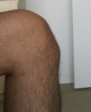
The Osgood-Schlatter disease is localized at the tibial tubercle, distal and anterior to the knee,
Clinical Presentation and Examination[edit | edit source]
Anterior knee pain associated with or without swelling, is the leading symptom in this disease and it aggravates during physical activities such as running, jumping, cycling, kneeling, walking up and down the stairs and kicking a ball(knee extension) . The clinical picture consists of pain localized to the area of the tibial tubercle.
- Painful palpation of the tibial tuberosity.
- Pain at the tibial tuberosity that worsens with physical activity or sport.
- Increased pain at the tibial tuberosity with sports activity.
- In some cases increased bony protuberance at the tibial tuberosity.
- Secondary to pain,Ely's test- there is tightness of Quadriceps.
- Resisted isometrics of the Quadricep muscle is painful.[1]
Some extra information and excursions available in this video
Differential Diagnosis[edit | edit source]
Conditions that show similar presentation:
- Jumper’s knee (patellar tendinitis) or Sinding- Larsen-Johanssen syndrome
- Synovial plica injury
- Tibial tubercle fracture
- Osteochondroma of the proximal tibia.
These diseases are also localized at the patellar tendon and can cause similar knee problems.
Diagnostic Procedures[edit | edit source]
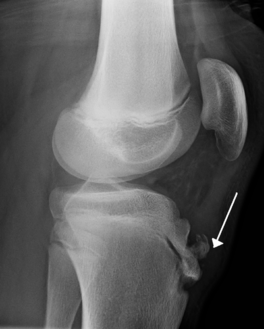
Radiological imagining is usually not required, and is done in severe cases or if an avulsion is suspected.
- The diagnosis is based on typical clinical findings (see clinical presentation). [3]
- Radiographic examinations of both knees should always be performed, in both the anterior-posterior and lateral projections, to rule out the possibility of tumors, fractures, ruptures or infections. A picture of prominent tibial tubercle with soft tissue swelling, calcification of the patellar tendon, or free bony fragment proximal to the tubercle can be seen.
- An utmost care must be taken, for accurate diagnosis, as sometimes the tibial protuberance may not be pathological. Therefore, clinical correlation must be done.
Prognosis[edit | edit source]
Excellent prognosis.[1]
The condition is self-limiting and hence recovers within a month
Sometimes the pain may persist up to 2 years, if unnoticed or left untreated.
Medical Management[edit | edit source]
Treatment should begin with rest, icing (RICE), activity modification and sometimes non-steroidal anti-inflammatory drugs.
Physical Therapy Management[edit | edit source]
Exercise Therapy[edit | edit source]
The physiotherapist can focus on exercises to improve the flexibility and strenghten the surrounding musculature. This includes the quadriceps, hamstring, iliotibial band and the gastrocnemius muscle. High-intensity quadriceps-strengthening exercises increase stress across the tibial tuberosity and are initially avoided. Stretching should initially be performed statically at a low intensity to prevent pain before progressing to dynamic or PNF stretching. A duration of at least thirty seconds with three repetitions is recommended at least once a day to increase the range of motion. Low-intensity quadriceps-strengthening exercises, such as isometric multiple- angle quadriceps exercises, are therefore instituted earlier in the conditioning program. High-intensity quadriceps exercises and hamstring stretching are introduced gradually and have been proven effective with high evidence rating. (level of evidence: 1a) [4]
Shockwave[edit | edit source]
Extracorporeal Shockwave therapy is a treatment which has been discussed in the use of osgood-schlatters but due to the low value evidence reccomendations cannot be made for this treatment. (level of evidence: 4) [5]
Activity Limitation[edit | edit source]
Non-operative treatment of this disease is based on the same principles that apply all overuse injuries. Today, there is no need for total immobilization, or for totally refraining from athletic activities. Of vital importance is that the physician inform the parents, the coach, and the child athlete of the natural course of this disease. The child should continue his normal physical activities, to the limit that the pain allows it, so lower intensity of frequency of exercising (activity modification). Also swimming, as a secondary athletic activity, is very good during this disease (no discomfort). Also knee-braces, tapes, slip-on knee support with an infrapatellar strap or pad are recommended and may help during physical activities and can reduce pain. (level of evidence: 5) [6]
Research by Gerulis et al (level of evidence: 2b) [7] has shown that limitation of physical activity, physical load restriction, and conservative treatment are more effective than physical load restriction and activity limitation alone
Taping[edit | edit source]
Taping: patellar tendon unloading technique
This technique has not been researched in this patient’s population; however, patients report that it decreases the pain. There are different usable ways to perform this technique.
The effectiveness of McConnell Taping has not been researched for this patient population; however, use of this taping technique alone has been attributed to improved knee flexion range of motion and decreased pain with activity. (level of evidence: 2b)[8] .
Surgical treatment[edit | edit source]
Surgical procedures should be avoided until the child has grown up and the bone growth has been completed to avoid growth-plate arrest and the development of recurvatum and or valgus of the knee. Surgical treatment, we identified different surgical procedures such as drilling of the tibial tubercle, excision of the tibial tubercle (decreasing the size), longitudinal incision in the patellar tendon, excision of the ununited ossicle and free cartilaginous pieces (tibial sequesstrectomy), insertion of bone pegs and/or a combination of any of these procedures.
References[edit | edit source]
- ↑ 1.0 1.1 1.2 Vaishya R, Azizi AT, Agarwal AK, Vijay V. Apophysitis of the tibial tuberosity (Osgood-Schlatter Disease): a review. Cureus. 2016 Sep;8(9).
- ↑ Baltaci G, Ozer H, Tunay V. B. Rehabilitation of avulsion fracture of the tibial tuberosity following Osgood-Schlatter disease. © Springer-Verlag 2003, Knee Surg Sports Traumatol ArthroscfckLR(2004) 12 : 115–118 www.springerlink.com/content/xnbxdt65aggv30dy/fulltext.pdffckLRLevel of evidence 4
- ↑ Çakmak S , Tekin L, Akarsu S. Long-term outcome of Osgood-Schlatter disease: not always favorable. Rheumatology International January 2014, Volume 34, Issue 1, pp 135-136
- ↑ Purushottam A. Gholvea, David M. Schera,b, Saurabh Khakhariaa,Roger F. Widmanna,b and Daniel W. Greena,b. Osgood Schlatter syndrome. Curr Opin Pediatr 19:44–50.2007 Lippincott Williams & Wilkins. Level Of Evidence : 1A
- ↑ Sportverletz Sportschaden. 2012 Dec;26(4):218-22. doi: 10.1055/s-0032-1325478. Epub 2012 Oct 9.[Extracorporeal shock wave therapy for patients suffering from recalcitrant Osgood-Schlatter disease].Lohrer H, Nauck T, Schöll J, Zwerver J, Malliaropoulos N. Level Of Evidence : 4
- ↑ C Reid D. Sports injuries assessment and rehabilitation. Churchill Livingstone USA, 1992, p 406 – 411 (level of evidence: 5)
- ↑ Gerulis V, Kalesinskas R, Pranckevicius S, Birgeris P. Importance of conservative treatment and physical load restriction to the course of osgood-schlatter's disease. Clinic of Pedia Surgery. 2003;2(5):57-64. Level Of Evidence : 2B
- ↑ Mason M, Keays SL, Newcombe PA. The effect of taping, quadriceps strengthening, and stretching prescribed separately or combined on patellofemoral pain. Physiother. 2011;16:109-119. Level Of Evidence : 2B


