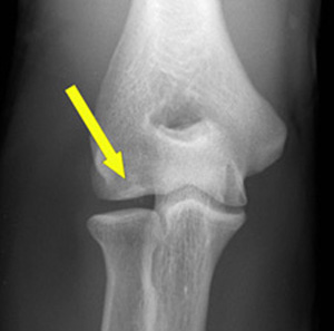
Osteochondritis Dissecans of the Elbow
Definition/Description [edit | edit source]
Osteochondritis Dissecans (OCD) is defined as an inflammatory pathology of bone and cartilage.This can result in localised necrosis and fragmentation of bone and cartilage.

OCD of the elbow is most commonly seen in the sporting adolescent population (ages 12-14) in particular throwing sports or upper limb dominant sports such as baseball or hockey.[2][3] Hence the common term "Little league elbow".[4]
In the elbow, the most common area affected is the capitellum, although it has been reported to affect the olecranon and the trochlea.[5][3] OCD can mean one or more flakes of articular cartilage have become separated. Which form loose bodies within the joint. The separated flakes can then ossify due to nourishment by the synovial fluid.[6] The cartilage is damaged and can form a loose body.[7]
In the long term OCD can lead to subsequent degenerative arthritis or osteoarthritis.[2]
Clinically Relevant Anatomy[edit | edit source]
Involved anatomy of this disorder includes the radial head or the central and/or lateral aspect of the capitellum.
Most OCD lesions of the elbow involve the capitellum, typically the central or lateral portion, but also the radial head, the olecranon of the ulna and the trochlea humeri.[2]
Epidemiology / Aetiology[edit | edit source]
Ostechondritis of the humeral capitellum is secondary to repetitive compression forces between radial head and capitellum.
Repetitive high stress forces on the joint can result in a series of minor injuries on the elbow that can eventually lead to bony fragmentation and ultimately detachment of the bony fragment from the bone.[3]
Commonly seen in the adolescent sporting population; who partake in repetitive throwing or overhead activities such as baseball and gymnastics.[7] More frequently seen in males (ages 10-14) than females and often affecting the dominant arm.[3][7]
Stages of osteochondritis dissecans:[5][edit | edit source]
Stage I[edit | edit source]
Thickening of cartilage and a stable lesion
Stage II[edit | edit source]
Articular cartilage interrupted and a stable lesion low signal rim behind fragment showing that there is fibrous attachment
Stage III[edit | edit source]
Articular cartilage interrupted, Unstable high signal changes behind fragment and underlying subchondral bone
Stage IV[edit | edit source]
Loose body Unstable
The cause of OCD is likely multifactorial. Causes of this pathology normally include injury or repetitive stress on the joint, lack of blood supply, and/or genetic makeup[5].
Some other mechanisms that can contribute to the development of OCD are: trauma, ischaemia, disordered ossification and genetic abnormalities. However, these mechanisms are not universally accepted but may be a contributing factor.[2]
Vascular hypo perfusion and repeated microtrauma may also contribute to the development of OCD. Capillary blood supply is often limited to 1 or 2 end vessels with limited collateral flow. This leads to vascular hypo perfusion.
Repeated microtrauma could lead to a production of a relatively avascular state in the vulnerable immature capitellar chondroepiphysis.[2]
Characteristics/Clinical Presentation[edit | edit source]
Presentation includes[3]:
- Lateral Pain over the joint
- Stiffness
- Feeling of instability
- Stiffness after resting
- Locking
- Giving way
- Popping/clicking
Differential Diagnosis[edit | edit source]
If there is no radiological confirmation of Osteochondritis Dissecans, other diagnoses may include:
- Panner's Disease in younger Shildren (9-10 years)[3]
- Insertional Apophysitis in pre-pubescent patients[4]
- Rheumatoid arthritis
- Osteoarthritis[2]
- Bone cysts
- Septic arthritis
- Epicondylar avulsion fractures in older patients[4]
Diagnostic Procedures [edit | edit source]
Radiographs can detect any abnormalities on the surface of the joint. Radiographs and MRI can also confirm diagnosis, monitor progress and assess for potential surgical intervention.[8]
X-ray can show if the cartilage flake includes a piece of underlying bone.[5]
Magnetic Resonance Imaging (MRI) will show any accumulation of fluid in the area and can detect any loose fragments. MRI should be considered when symptomatic patients have a normal X-Ray.[9]
Computerised Tomography (CT) can detect any bony fragments, pinpoint their location and determine whether they have settled in the joint space.[10]
Outcome Measures [edit | edit source]
An outcome measure appropriate for this injury is the DASH questionnaire which measures disabilities of the elbow, shoulder and hand as they relate to everyday function.[11][12]Additional measures may be required in terms of return to sport, especially in professional level sport.
Examination[edit | edit source]
Physical examination is possible to find radiocapitellar tenderness. Crepitus, decreased elbow extension and an effusion could be occurring. As told above clicking, catching or locking could also occur. These suggest OCD fragment instability and the associated loose bodies. To help confirm the diagnosis of capitellar OCD we can use the active radiocapitellar compression test. The test goes as follows: the examiner places the elbow of the patient in full extension and then he lets the patient actively pronate and supinate the forearm. (Level of Evidence 3A) This test is positive if it results in a compression across the radiocapitellar joint and which evokes lateral joint pain.[2]
Medical Management [edit | edit source]
Surgical management may be an option if conservative care fails or if the lesion is Grade III or higher.
Surgical interventions are aimed at:
Promoting re-vascularisation to the area
Debriding the area
Fragment stabilisation
Bone grafting if needed
Most of these procedures are done arthroscopically (minimally invasive via portal sites), although depending on the size of the loose fragments an open procedure may be required.
Operative Management
The mainstay of the operative management is the removal of the loose fragments and debridement of the base, also the arthroscopic evaluation of the lesion is a part of the treatment. Although most of the older literature was focused on an open management, most of the newest literature delineate the efficacy of arthroscopic surgery. The diagnostic arthroscopy of the anterior compartment of the elbow is the first step for the operative technique (Level of Evidence 2A). The most useful technique is to begin the arthroscopy with the anteromedial portal to visualise the radiocapitellar joint. The anterior part of the capitulum is mostly normal. If we notified a anterior lesion, we can use the 70° arthroscope to visualise the lesion and the lateral portal is used to treat the lesion. The anterior side of the elbow is then evaluated with a camera for loose bodies. If there are loose bodies, they are removed via an anterolateral portal. Also the fossa olecranon is evaluated for loose bodies. If there are loose bodies, they are removed via a posterior central or posterior lateral portal. The medial gutter is also evaluated for loose bodies and inflammation. If there is any inflammation or thickening of the posterolateral plica, they remove it via a posterolateral portal.[3]
Arthroscopic surgery can include debridement, fragment fixation, microfracturing and osteochondral autografting. Removing of the fragments alone is a minimally and common invasive procedure. The long-term results can be poor if a large osteochondral defect of the capitulum remains. An alternative procedure is the fragment fixation with a bone graft. It is difficult to fix a cartilaginous fragment securely in unhealthy subchondral bone (Level of Evidence 3A). Recurrent symptoms after a short time span can be expected in these patients. Research show us a gain in ROM about debridement, microfracturing, drilling and creating fibrocartilage. The hyaline cartilage can mostly not be restored with the techniques of treatment of capitellar OCD. The technique that can restore the hyaline cartilage is the osteochondral autografting technique. The negative point of osteochondral autografting is the need to expose the knee joint to fix the graft. But for the elbow the size of the defect is limited. [13]
Physical Therapy Management[edit | edit source]
Conservative care for this condition include physical therapy, use of non-steroidal anti-inflammatory drugs (NSAIDs), rest from sport for 6-8 weeks, and bracing.
Steven A. Giuseffi states that the non-operative treatment of Osteochondritis Dissecans of the elbow still remains controversial. Lesion stability, integrity of the overlying cartilage and status of the capitellar physis are key considerations in determining the appropriate management. Lesions with an intact cartilage surface, i.e. a stable lesion, but also non-displaced lesions with cartilage compromise can be treated using a non-operative management. If an open physis, relatively preserved elbow range of motion (ROM), 20° at most, localised flattening or lucency on radiographs without evidence of defect fragmentation or instability occurs a positive prognosis could be stated. [2]
This non-operative treatment consists of rest and activity modification, as such that sports and aggravating activities are ceased until symptoms resolve. This usually takes 3 to 12 weeks until the symptoms are resolved.
Steven A. Giuseffi also recommends the use of a hinged brace during the passing time. This protects the elbow and helps correct a natural elbow valgus and will help offload the capitellum. When the patient’s symptoms improve, gentle physical therapy and ROM-exercises could be used. At first, the brace is set at the limits of pain-free range of motion, often as limited as 60 to 90°. The brace is loosened to allow full range of motion, as the inflammation in the plica decreases and pain-free motion increases (Level of Evidence 2A), [3] Within 2 weeks from the beginning of treatment, the patient is able to return to normal activities with the brace in place. [3] After 3 to 6 months (usually) athletes can return to their sports activities.The patient and family should be advised of potential long-term sequelae.
If the patient doesn’t have a stable lesion, operative treatment could be advised. If arthroscopic debridement and drilling was used, non-operative treatment could be used postoperative. The patient will be placed in a double-hinged elbow brace within a week and gentle ROM-exercises could be used. When pain and swelling disappear, the patient may gradually participate in athletic activities in the brace. Full return to athletics could be estimated after 3-4 months. (Level of Evidence 3A) As mentioned above patients and parents should be advised of potential sequelae. [2]
The patient can be situated in three phases: acute, recovery and maintenance.
During the acute phase the physiotherapist should advise the patient, who have an intact humeral capitellum osteochondritis dissecans lesion, to rest and to limit their activities. He/she should also teach these patients how to apply ice on their elbow. If the patient experiences a lot of pain, the physiotherapist should consider using a hinge brace to relieve this pain and only for this use. Bracing during a long period of time is not indicated as these could increase the risk of flexion contractures. As referred before it’s very important to apply ice, as this relieves the pain. [14]
When the patient could be located in the recovery phase, after an intervention, then the physiotherapist can start with passive range-of-motion (ROM) exercises. These can be follow by active ROM exercises, this to avoid contractures. Progressive resistance training should be used as the patient can tolerate more activity and gets stronger. It hasn’t been proven in CT’s (clinical trials) but electrical stimulation may also facilitate recovery. [14]
The maintenance phase consists of continued use of strengthening and ROM exercises for the elbow. It is also important to prevent further injuries by taking measures protecting the elbow. The physiotherapist could advise patients of the significance of a wrist/finger program and a shoulder program. Strengthening and ROM exercises should also be the main focus during these programs. [14]
Analytic physiotherapy consists of specific techniques. Those techniques could be used for traumatic osteoarticular pathologies, micro-traumas, functional or postural pathologies. This states that it could be used for inflammatory pathologies as osteochondritis.
The treatment starts with specific mobilisation meaning to release blocked and displaced joints. These mobilisations are calm and progressive. These movements are executed in the initial position, this to evade tension on soft tissue. Researchers express on the demanded cyclic force that should be given. The cyclic force will lead to a normal physiology of chondrocytes. Constant pressure will result in destruction of the cartilage. Switching up the contraction and relaxation of muscles during these techniques is also important. Following these steps will result in regeneration of the articular structures to an optimal state. [15]
Key Evidence[edit | edit source]
A study published in 2008 reports a favorable prognosis of up to 90% for patients who are treated conservatively in the stage I of this condition. Patients who were in stage II had about a 53% of a good prognosis with conservative treatment. Duration of this treatment was just under 15 months in the first stage and 13 months in the second stage. About 78% of stage I and 53% of stage II patients were able to return to competitive baseball. Mean ages for these groups were about 12 years in stage I and 14 years of age in stage II. This study is a level III study, and more research should be done to support these results.
Another study performed in 2006 looks at the surgical considerations for osteochondritis dissecans. The article looks at 15 patients with a mean age of 28 that underwent elbow arthroscopic debridement for this condition. These patients were classified using the Baumgarten system and were graded during arthroscopy. They looked at the patients about 4 years out of surgery and found a significant decrease in pain levels and function was improved from a “poor” to an “excellent” status per the modified Andrews elbow scoring system. About 80% of these patients were able to return to the same level of sport while all of the patients were able to return to work.
Factors that should be considered are age, activity level for the studies above. Clearly more research needs to be done to determine best practice when it comes to treatment of osteochondritis dissecans.
Resources[edit | edit source]
Resources we used for pictures:
Fig 1 : https://www.youtube.com/watch?v=sfAxCsv_Hes (12)
Fig. 2: http://radiology.casereports.net/index.php/rcr/article/view/18/161 (11)
Fig. 3: http://www.seattlechildrens.org/medical-conditions/bone-joint-muscle-conditions/cartilage-conditions/osteochondritis-dissecans/ (16)
Fig. 4: http://www.boneclinic.com.sg/patient-education/child-orthopaedics/adolescent-osteochondritis-dissecans/ (17)
Books:
H.L.F. Currey,Essentials of rheumatology, 1988, p166 [9]
Clinical Bottom Line [edit | edit source]
Osteochondritis Dissecans [2] is, in adolescent athletes, an increasingly common cause of elbow dysfunction and elbow pain. [6] It can lead to additional conditions which lead to a complex situation. There are multiple mechanisms that lead to OCD: trauma, ischemia, disordered ossification and genetic abnormalities. What typically happens is that one or more flakes of articular cartilage separates and forms loose bodies within the joint. [2] The separated flakes remain alive and frequently ossify. [14] Most lesions involve the capitellum, typically the central or lateral portion, but also the radial head, the olecranon of the ulnae and the trochlea humeri. [3] OCD can also be caused by a vascular dysfunction or microtrauma. [2] Main characteristics of the illness that are commonly mentioned are locking, giving away and recurrent effusions. [3] As far as the differential diagnosis goes, we can quote Panner’s disease, osteonecrosis, osteochondral fracture, hereditary epiphyseal dysplasia and Little Leaguer’s Elbow. [2] Examination can be done using the active radiocapitellar compression test, in case of lateral joint pain evocation the test is considered positive. [2] For medical management, the use of arthroscopic surgery is one of the options. [3] As for the physical therapy management it still remains controversial, although there is a vast description of the possibilities described. [2]
References [16] [17][18] [19] [20][edit | edit source]
- ↑ Folio LR, Craig SH, Wright GA, Battaglia MJ. Loose body in elbow of a baseball player: arthroscopic/radiologic correlation. Radiology case reports. 2006 Jan 1;1(2):62-5.
- ↑ 2.00 2.01 2.02 2.03 2.04 2.05 2.06 2.07 2.08 2.09 2.10 2.11 2.12 2.13 2.14 Giuseffi SA, Field LD. Osteochondritis dissecans of the elbow. Operative Techniques in Sports Medicine. 2014 Jun 1;22(2):148-55.
- ↑ 3.00 3.01 3.02 3.03 3.04 3.05 3.06 3.07 3.08 3.09 3.10 3.11 Felix H. Savoie, III, MD. Osteochondritis Dissecans of the Elbow. Operative Techniques in Sports Medicine. 2008 16:187-193
- ↑ 4.0 4.1 4.2 Klingele KE, Kocher MS. Little league elbow. Sports Medicine. 2002 Dec 1;32(15):1005-15.
- ↑ 5.0 5.1 5.2 5.3 Baker III CL, Romeo AA, Baker Jr CL. Osteochondritis dissecans of the capitellum. The American journal of sports medicine. 2010 Sep;38(9):1917-28.
- ↑ Curry H. Essentials of Rheumatology. 1st ed. Churchill Livingstone; 1988.
- ↑ 7.0 7.1 7.2 Matsuura T, Suzue N, Iwame T, Nishio S, Sairyo K. Prevalence of osteochondritis dissecans of the capitellum in young baseball players: results based on ultrasonographic findings. Orthopaedic journal of sports medicine. 2014 Aug 11;2(8):2325967114545298.
- ↑ Zbojniewicz AM, Laor T. Imaging of osteochondritis dissecans. Clinics in sports medicine. 2014 Apr 1;33(2):221-50.
- ↑ Brunton LM, Anderson MW, Pannunzio ME, Khanna AJ, Chhabra AB. Magnetic resonance imaging of the elbow: update on current techniques and indications. The Journal of hand surgery. 2006 Jul 1;31(6):1001-11.
- ↑ Moktassi A, Popkin CA, White LM, Murnaghan ML. Imaging of osteochondritis dissecans. Orthopedic Clinics. 2012 Apr 1;43(2):201-11.
- ↑ Schoch B, Wolf BR. Osteochondritis dissecans of the capitellum: minimum 1-year follow-up after arthroscopic debridement. Arthroscopy: The Journal of Arthroscopic & Related Surgery. 2010 Nov 1;26(11):1469-73.
- ↑ MacDermid JC. Outcome evaluation in patients with elbow pathology: issues in instrument development and evaluation. Journal of Hand Therapy. 2001 Apr 1;14(2):105-14.
- ↑ Frank de Graaff et al., Arthroscopic Surgery in Athletes With Osteochondritis Dissecans of the Elbow, Artrhoscopy, 2011 (Level of Evidence : 3A)
- ↑ 14.0 14.1 14.2 14.3 Shital Patel. Humeral Capitellum Osteochondritis Dissecans Treatment & Management, MedScape. (Level of evidence: 3A)
- ↑ Michel Haye, La kinésithérapie analytique thérapie manuelle selon le concept de Sohier,Profession Kinésithérapeute, 2007 (Level of evidence: 3A)
- ↑ Mayo Clinic. Osteochondritis Dissecans. http://www.mayoclinic.com/health/osteochondritis-dissecans/DS00741 (accessed 6/13/09).
- ↑ Rahusen F Th G, et al. Results of arthroscopic debridement for osteochondritis dissecans of the elbow. B J Sports Med 2006;40:966-969. Abstract: http://bjsportmed.com/cgi/content/abstract/40/12/966 (accessed 6/25/09).
- ↑ Matsuura T, et al. Conservative Treatment for Osteochondrosis of the Humeral Capitellum. Am J Sports Med. 2008;36(5): 868-872. Abstract: http://ajs.sagepub.com/content/36/5/868.abstract (accessed 6/18/09).
- ↑ O’Sullivan S, Siegelman R. National Physical Therapy Examination Review & Study Guide. Evanston, IL: International Educational Resources Ltd, 2006.
- ↑ Patient UK. http://www.patient.co.uk/showdoc/40001133/ (accessed 6/13/09)


