Pelvis: Difference between revisions
Nicole Hills (talk | contribs) No edit summary |
Nicole Hills (talk | contribs) mNo edit summary |
||
| Line 1: | Line 1: | ||
<div class="noeditbox">This page is currently undergoing work, but please come back later to check out new information</div> | <div class="noeditbox">This page is currently undergoing work, but please come back later to check out new information</div> | ||
== Description == | == Description == | ||
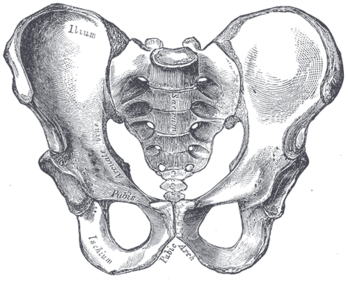
[[File:Gray241.png|alt=Bones of the pelvis|thumb| | [[File:Gray241.png|alt=Bones of the pelvis|thumb|348x348px]]The pelvis consists of the sacrum, the coccyx,the ischium, the ilium, and the pubis. <ref name=":1">White, TD., Black, MT., Folkens, PA. Human osteology. Academic press; 2011.</ref><ref name=":0">Lewis CL, Laudicina NM, Khuu A, Loverro KL. The human pelvis: Variation in structure and function during gait. The Anatomical Record. 2017 Apr;300(4):633-42.</ref> The structure of the pelvis supports the contents of the abdomen while also helping to transfer the weight from the spine to the lower limbs.<ref name=":2">Magee DJ. Orthopedic physical assessment. Elsevier Health Sciences; 2013 Dec 4.</ref> During gait, the joints within the pelvis work together to decrease the amount of force transferred from the ground and lower extremities to the spine and upper extremities.<ref name=":2" /> | ||
== Anatomy == | == Anatomy == | ||
| Line 12: | Line 10: | ||
** ischium | ** ischium | ||
** ilium | ** ilium | ||
** pubis | ** pubis<ref name=":1" /> | ||
=== Joint Articulations === | |||
=== Articulations === | |||
There are three articulations within the pelvis: | There are three articulations within the pelvis: | ||
* inferiorly between the sacrum and the coccyx | * inferiorly between the sacrum and the coccyx | ||
* posteriorly between the sacrum and each ilium ([[Sacroiliac joint|sacroiliac (SI) joint]]) | * posteriorly between the sacrum and each ilium ([[Sacroiliac joint|sacroiliac (SI) joint]]) | ||
* anteriorly between the pubic bodies (pubic symphysis).<ref name=":0" /> | * anteriorly between the pubic bodies (pubic symphysis).<ref name=":0" /> | ||
Other articulations: | |||
= | * the pelvis and femur articulate via the acetabulum<ref name=":1" /> | ||
=== Ligaments === | === Ligaments === | ||
=== Muscles === | === Muscles === | ||
There are 35 muscles that attach to the sacrum or innominates which mainly provide stability to the joint rather than producing movements.<ref>Calvillo O., Skaribas I., Turnispeed J., Anatomy and pathophysiology of the SIJ, current science, 2000 (LOE 2A)</ref> | |||
Muscles that attach to the sacrum or innominates: | |||
* Adductor brevis | |||
* Adductor longus | |||
* [[Adductor Magnus|Adductor magnus]] | |||
* [[Biceps Femoris|Biceps femoris - long head]] | |||
* [[Pelvic Floor Anatomy|Coccygeus]] | |||
* [[Erector spinae]] | |||
* [[Abdominal Muscle Anatomy|External oblique]] | |||
* [http://www.rad.washington.edu/academics/academic-sections/msk/muscle-atlas/lower-body/gluteus-maximus][[Gluteus Maximus|Gluteus maxiumus]] | |||
* [http://www.rad.washington.edu/academics/academic-sections/msk/muscle-atlas/lower-body/gluteus-medius][[Gluteus Medius|Gluteus medius]] | |||
* [[Gluteus Minimus|Gluteus minimus]] | |||
* Gracilis | |||
* [http://www.rad.washington.edu/academics/academic-sections/msk/muscle-atlas/lower-body/psoas][[Iliacus]] | |||
* Inferior gemellus | |||
* [[Abdominal Muscle Anatomy|Internal oblique]] | |||
* [http://www.rad.washington.edu/academics/academic-sections/msk/muscle-atlas/upper-body/latissimus-dorsi][[Latissimus Dorsi Muscle|Latissimus dorsi]] | |||
* [[Pelvic Floor Anatomy|Levator ani]] | |||
* Multifidus | |||
* Obturator internus | |||
* Obturator externus | |||
* Pectineus | |||
* [[Piriformis]] | |||
* [[Psoas Minor|Psoas minor]] | |||
* Pyramidalis | |||
* Quadratus femoris | |||
* [[Quadratus Lumborum|Quadratus lumborum]] | |||
* [[Rectus Abdominis|Rectus abdominis]] | |||
* Rectus femoris | |||
* Sartorius | |||
* Semimembranosus | |||
* Semitendonosus | |||
* [[Pelvic Floor Anatomy|Sphincter urethrae]] | |||
* [[Pelvic Floor Anatomy|Superficial transverse perineal ischiocavernous]] | |||
* Superior gemellus | |||
* [[Tensor Fascia Lata|Tensor fascia lata]] | |||
* [[Abdominal Muscle Anatomy|Transversus abdominus]] | |||
=== | === Sex-specific differences === | ||
== Clinical Examination == | == Clinical Examination == | ||
=== Assessment === | === Assessment === | ||
* | *Prior to the assessment of the sacroiliac joint both the lumbar spine and hip should be assessed and any underlying pathologist should be ruled out. | ||
=== Special Tests === | === Special Tests === | ||
**[[ | |||
** | ==== SI Joint Stress tests ==== | ||
**[[ | * Anterior Gapping test | ||
** | * [[Sacroiliac Distraction Test|Sacroiliac Distraction test]] | ||
** | * Pubic Stress test | ||
**[[ | * Sacrotuberous Ligament Stress test | ||
** | * Sacral Compression test | ||
**[[FABER Test|FABER | * Rotational Stress test | ||
**[[ | |||
==== [[Leg Length Discrepancy|Leg Length tests]] ==== | |||
* Prone test | |||
* Standing leg length test | |||
* Functional leg length test | |||
==== Other Special Tests ==== | |||
* Seated Flexion test (Piedallu's Sign) | |||
* Long Sit test | |||
* [[Sign of the Buttock]] | |||
* Posterior Pelvic Pain Provocation test | |||
* Gaenslen's test | |||
* Yeoman's test | |||
* [[FABER Test|FABER (Figure-Four) test]] | |||
* Fortin Finger Test | |||
* [[Straight Leg Raise Test|Straight Leg Raise - 70-90deg]] | |||
* Gillet's test (Ipsilateral posterior rotation test) | |||
=== Outcome Measures === | === Outcome Measures === | ||
Revision as of 17:21, 5 September 2018
Description[edit | edit source]
The pelvis consists of the sacrum, the coccyx,the ischium, the ilium, and the pubis. [1][2] The structure of the pelvis supports the contents of the abdomen while also helping to transfer the weight from the spine to the lower limbs.[3] During gait, the joints within the pelvis work together to decrease the amount of force transferred from the ground and lower extremities to the spine and upper extremities.[3]
Anatomy[edit | edit source]
Osteology[edit | edit source]
- sacrum
- coccyx
- two innominate bones, which consist of the:
- ischium
- ilium
- pubis[1]
Joint Articulations[edit | edit source]
There are three articulations within the pelvis:
- inferiorly between the sacrum and the coccyx
- posteriorly between the sacrum and each ilium (sacroiliac (SI) joint)
- anteriorly between the pubic bodies (pubic symphysis).[2]
Other articulations:
- the pelvis and femur articulate via the acetabulum[1]
Ligaments[edit | edit source]
Muscles[edit | edit source]
There are 35 muscles that attach to the sacrum or innominates which mainly provide stability to the joint rather than producing movements.[4]
Muscles that attach to the sacrum or innominates:
- Adductor brevis
- Adductor longus
- Adductor magnus
- Biceps femoris - long head
- Coccygeus
- Erector spinae
- External oblique
- [1]Gluteus maxiumus
- [2]Gluteus medius
- Gluteus minimus
- Gracilis
- [3]Iliacus
- Inferior gemellus
- Internal oblique
- [4]Latissimus dorsi
- Levator ani
- Multifidus
- Obturator internus
- Obturator externus
- Pectineus
- Piriformis
- Psoas minor
- Pyramidalis
- Quadratus femoris
- Quadratus lumborum
- Rectus abdominis
- Rectus femoris
- Sartorius
- Semimembranosus
- Semitendonosus
- Sphincter urethrae
- Superficial transverse perineal ischiocavernous
- Superior gemellus
- Tensor fascia lata
- Transversus abdominus
Sex-specific differences[edit | edit source]
Clinical Examination[edit | edit source]
Assessment[edit | edit source]
- Prior to the assessment of the sacroiliac joint both the lumbar spine and hip should be assessed and any underlying pathologist should be ruled out.
Special Tests[edit | edit source]
SI Joint Stress tests[edit | edit source]
- Anterior Gapping test
- Sacroiliac Distraction test
- Pubic Stress test
- Sacrotuberous Ligament Stress test
- Sacral Compression test
- Rotational Stress test
Leg Length tests[edit | edit source]
- Prone test
- Standing leg length test
- Functional leg length test
Other Special Tests[edit | edit source]
- Seated Flexion test (Piedallu's Sign)
- Long Sit test
- Sign of the Buttock
- Posterior Pelvic Pain Provocation test
- Gaenslen's test
- Yeoman's test
- FABER (Figure-Four) test
- Fortin Finger Test
- Straight Leg Raise - 70-90deg
- Gillet's test (Ipsilateral posterior rotation test)
Outcome Measures[edit | edit source]
Pathology/Injury[edit | edit source]
- Pubic Symphysis Dysfunction
- Spondyloarthropathies
- Pregnancy Related Pelvic Pain
- Pelvic Fractures
- Sacroiliitis
Physiotherapeutic Techniques[edit | edit source]
Resources[edit | edit source]
- ↑ 1.0 1.1 1.2 White, TD., Black, MT., Folkens, PA. Human osteology. Academic press; 2011.
- ↑ 2.0 2.1 Lewis CL, Laudicina NM, Khuu A, Loverro KL. The human pelvis: Variation in structure and function during gait. The Anatomical Record. 2017 Apr;300(4):633-42.
- ↑ 3.0 3.1 Magee DJ. Orthopedic physical assessment. Elsevier Health Sciences; 2013 Dec 4.
- ↑ Calvillo O., Skaribas I., Turnispeed J., Anatomy and pathophysiology of the SIJ, current science, 2000 (LOE 2A)


