Popliteal Fossa: Difference between revisions
(hyper links) |
(hyperlinks) |
||
| Line 8: | Line 8: | ||
[[File:Popliteal_picture.jpg|center|frameless]] | [[File:Popliteal_picture.jpg|center|frameless]] | ||
=== Description === | === Description === | ||
The Popliteal Fossa is a diamond- shaped space behind the [[knee | The Popliteal Fossa is a diamond- shaped space behind the [[knee]] joint .<ref name=":1">Chummy,SS. Last's Anatomy.Twelfth Edition.Churchill Livingstone.2011.pg 132</ref> It is formed between the muscles in the posterior compartments of the thigh and leg.This anatomical landmark is the major route by which structures pass between the thigh and leg.<ref name=":0">Richard,LD, Wayne,VA,Adam,WM.Grays'Anatomy for Students. Second Edition.Churchill Livingstone.2010.pg 584-585</ref> | ||
=== Margins === | === Margins === | ||
The Popliteal Fossa has 2 upper margins and 2 lower margins<ref name=":0" /> | The Popliteal Fossa has 2 upper margins and 2 lower margins<ref name=":0" /> | ||
* The margins of the upper part are formed by the Semimembranosus and Semitendinosus muscles on the medial side and the Biceps Femoris on the lateral side. | * The margins of the upper part are formed by the [[Semimembranosus]] and [[Semitendinosus]] muscles on the medial side and the [[Biceps Femoris]] on the lateral side. | ||
* The margins of the lower parts are formed by the medial head of the Gastrocnemius muscle and the laterally by the Plantaris muscle and the lateral head of the Gastrocnemius muscle. | * The margins of the lower parts are formed by the medial head of the [[Gastrocnemius]] muscle and the laterally by the [[Plantaris]] muscle and the lateral head of the [[Gastrocnemius]] muscle. | ||
=== Floor === | === Floor === | ||
The floor of the fossa is formed by the Popliteal surface of the | The floor of the fossa is formed by the Popliteal surface of the [[Femur]], Capsule of the [[Knee]] reinforced by the oblique Popliteal [[ligament]] and the Popliteus muscle covered by its [[Fascia]]<ref name=":1" />. | ||
=== Roof === | === Roof === | ||
The roof of the Popliteal fossa is covered by the Fascia Lata which is strongly reinforced by tranverse fibers. The roof is pierced by the small | The roof of the Popliteal fossa is covered by the [[Tensor Fascia Lata|Fascia Lata]] which is strongly reinforced by tranverse fibers. The roof is pierced by the small Saphenous vein and the posterior Femoral cutaneous nerve<ref name=":1" />. | ||
=== Content === | === Content === | ||
The major content of the Popliteal fossa are the Popliteal artery,the Popliteal vein, the Tibia and | The major content of the Popliteal fossa are the Popliteal artery,the Popliteal vein, the [[Tibial Nerve|Tibia nerve]] and Common Fibular nerve. <div class="researchbox"> </div> | ||
=== Clinical Significance === | === Clinical Significance === | ||
* | * [[Baker's Cyst|Baker's cyst]] | ||
* Popliteal Pulse | * Popliteal [[Pulse rate|Pulse]] | ||
* Popliteal Aneurysm | * Popliteal Aneurysm | ||
Revision as of 03:55, 23 September 2020
Original Editor - User:Ochia Lilian Chidera
Top Contributors - Ochia Lilian Chidera, Kirenga Bamurange Liliane, Lucinda hampton and Kim Jackson
Description[edit | edit source]
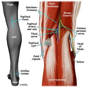
The Popliteal Fossa is a diamond- shaped space behind the knee joint .[1] It is formed between the muscles in the posterior compartments of the thigh and leg.This anatomical landmark is the major route by which structures pass between the thigh and leg.[2]
Margins[edit | edit source]
The Popliteal Fossa has 2 upper margins and 2 lower margins[2]
- The margins of the upper part are formed by the Semimembranosus and Semitendinosus muscles on the medial side and the Biceps Femoris on the lateral side.
- The margins of the lower parts are formed by the medial head of the Gastrocnemius muscle and the laterally by the Plantaris muscle and the lateral head of the Gastrocnemius muscle.
Floor[edit | edit source]
The floor of the fossa is formed by the Popliteal surface of the Femur, Capsule of the Knee reinforced by the oblique Popliteal ligament and the Popliteus muscle covered by its Fascia[1].
Roof[edit | edit source]
The roof of the Popliteal fossa is covered by the Fascia Lata which is strongly reinforced by tranverse fibers. The roof is pierced by the small Saphenous vein and the posterior Femoral cutaneous nerve[1].
Content[edit | edit source]
The major content of the Popliteal fossa are the Popliteal artery,the Popliteal vein, the Tibia nerve and Common Fibular nerve.Clinical Significance[edit | edit source]
- Baker's cyst
- Popliteal Pulse
- Popliteal Aneurysm


