Psoas Minor
Original Editor - Oyemi Sillo
Lead Editors - Kim Jackson, Eman Ammar, Lucinda hampton, Maram Salem, Oyemi Sillo and WikiSysop
Description[edit | edit source]
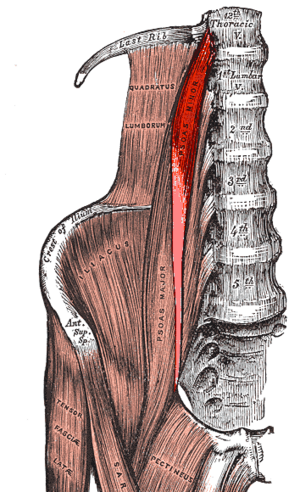
Psoas Minor is a thin paired muscle of the posterior abdominopelvic region, placed in front of the Psoas major.[1] The psoas minor muscle origininates from the last thoracic vertebra and the first lumbar; it is present in 60% to 65% of the population. Distally, it converges with the iliacus fascia and the psoas major tendon to insert on the iliopectineal eminence (for 90% of the population).
- The major and minor psoas muscles and the iliacus muscle make up the iliopsoas musculotendinous unit. Commonly called iliopsoas muscle. This complex muscle system can function as a unit or intervene as separate muscles. It is essential for correct standing or sitting lumbar posture, hip joint, and during walking and running[2].
Image 1: R Psoas Minor Highlighted in red
Anatomy[edit | edit source]
- Origin: Lateral aspect of vertebral body of 12th thoracic and 1st lumbar vertebrae[3]
- Insertion: Pectineal line of pubis[3]
- Nerve Supply: Small branch from the initial part of the lumbar ventral ramus(L1)[3]
- Blood Supply: Lumbar arteries, lumbar branch of the iliolumbar artery.[3]
Action[edit | edit source]
Assists with flexion of the lumbar vertebral column [3]
Physiotherapy[edit | edit source]
See
References[edit | edit source]
- ↑ Gray, Henry. Anatomy of the Human Body. Philadelphia: Lea & Febiger, 1918; Bartleby.com, 2000. www.bartleby.com/107/.
- ↑ Bordoni B, Varacallo M. Anatomy, Bony Pelvis and Lower Limb, Iliopsoas Muscle. StatPearls [Internet]. 2021 Jul 21. Available:https://www.ncbi.nlm.nih.gov/books/NBK531508/ (accessed 16.1.2022)
- ↑ 3.0 3.1 3.2 3.3 3.4 http://www.anatomyexpert.com/app/structure/5312/
- ↑ Anderson CN. Iliopsoas: pathology, diagnosis, and treatment. Clinics in sports medicine 2016;35(3):419-33. Micheo W. Musculoskeletal, Sports and Occupational Medicine. Demos Medical Publishing; 2010 Dec 21.


