Scapula: Difference between revisions
No edit summary |
No edit summary |
||
| Line 5: | Line 5: | ||
</div> | </div> | ||
== Osteology == | == Osteology == | ||
The scapula is a thin, flat triangular-shaped bone placed on the postero-lateral aspect of the thoracic cage. It has 2 surfaces, 3 borders, 3 angles and 3 processes. | |||
=== Surfaces === | === Surfaces === | ||
# <u>Costal Surface or Subscapular Fossa</u><br>It is concave and is directed medially and forwards. It is marked by 3 longitudinal ridges. A thick ridge adjoins the lateral border. This part of bone is almost rod like | # <u>Costal Surface or Subscapular Fossa</u><br>It is concave and is directed medially and forwards. It is marked by 3 longitudinal ridges. A thick ridge adjoins the lateral border. This part of the bone is almost rod-like acts a lever for the action of serratus anterior in overhead abduction of the arm. | ||
# <u>Dorsal Surface</u><br>The prominent spine of scapula divides the surface into a smaller | # <u>Dorsal Surface</u><br>The prominent spine of scapula divides the surface into a smaller supraspinous fossa and a larger infraspinous fossa. The depth of the supraspinous fossa is filled by the supraspinatus muscle. | ||
=== Borders === | === Borders === | ||
| Line 30: | Line 30: | ||
<br> | <br> | ||
{{#ev:youtube|fEXrPsGySKI|300}} <ref> Kenhub-Learn Human Anatomy.Anatomy and Function of the Scapula-Human Anatomy|Kenhub.Available from:https://youtu.be/fEXrPsGySKI (Assessed 31 July 2020) </ref> | {{#ev:youtube|fEXrPsGySKI|300}} <ref>Kenhub-Learn Human Anatomy.Anatomy and Function of the Scapula-Human Anatomy|Kenhub.Available from:https://youtu.be/fEXrPsGySKI (Assessed 31 July 2020) </ref> | ||
=== Attachments on Scapula === | === Attachments on Scapula === | ||
Scapula has various bony projections for attachment of muscles, ligaments and other soft-tissue structures.<ref name="WGR">Williams GR, Shakil M, Klimkiewicz J. Anatomy of the Scapulothoracic Articulation. Clin Orthop Relat Res. Feb 1999;237-46. [Medline].</ref> These include the scapular spine, acromion process, coracoid process, scapular notch, lateral scapular spine, and glenoid fossa. The suprascapular nerve travels through the notch and provides motor supply to the supraspinatus muscle and infraspinatus muscle, as well as sensation to the acromioclavicular joint.<ref name="WGR" /> <ref name="CE">Culham E, Peat M. Functional Anatomy of the Shoulder Complex. J Orthop Sports Phys Ther. Jul 1993;18(1):342-50. [Medline].</ref><br> | Scapula has various bony projections for attachment of muscles, ligaments and other soft-tissue structures.<ref name="WGR">Williams GR, Shakil M, Klimkiewicz J. Anatomy of the Scapulothoracic Articulation. Clin Orthop Relat Res. Feb 1999;237-46. [Medline].</ref> These include the scapular spine, acromion process, coracoid process, scapular notch, lateral scapular spine, and glenoid fossa. The suprascapular nerve travels through the notch and provides motor supply to the supraspinatus muscle and infraspinatus muscle, as well as sensation to the acromioclavicular joint.<ref name="WGR" /> <ref name="CE">Culham E, Peat M. Functional Anatomy of the Shoulder Complex. J Orthop Sports Phys Ther. Jul 1993;18(1):342-50. [Medline].</ref><br> | ||
=== Ossification === | === Ossification === | ||
The scapula ossifies from one primary centre and seven secondary centres. The primary centre appears near the glenoid cavity during the 8th week of development. The 1st secondary centre appears in the middle of coracoid process during the first year and fuses by the 15th year. The subcoracoid centre appears in the root of the coracoid process during the 10th year and fuses by the 16th to 18th years. The other centres, including 2 for the acromion , | The scapula ossifies from one primary centre and seven secondary centres. The primary centre appears near the glenoid cavity during the 8th week of development. The 1st secondary centre appears in the middle of coracoid process during the first year and fuses by the 15th year. The subcoracoid centre appears in the root of the coracoid process during the 10th year and fuses by the 16th to 18th years. The other centres, including 2 for the acromion , oe for the lower 2/3rds of the margin of the glenoid cavity, one for the medial border and one for the inferior angle, appear at puberty and fuse by the 25th year. <ref name="WGR" /> | ||
== Soft Tissue == | == Soft Tissue == | ||
| Line 42: | Line 42: | ||
* [[Subscapularis|Subscapularis]] arises from the medial 2/3rds of the subscapular fossa. | * [[Subscapularis|Subscapularis]] arises from the medial 2/3rds of the subscapular fossa. | ||
* [[Supraspinatus|Supraspinatus]] arises from medial 2/3rds of supraspinous fossa including upper surface of the spine | * [[Supraspinatus|Supraspinatus]] arises from medial 2/3rds of supraspinous fossa including upper surface of the spine | ||
* [[Infraspinatus|Infraspinatus arises]] from medial 2/3rds of infraspinous fossa, including lower surface of | * [[Infraspinatus|Infraspinatus arises]] from medial 2/3rds of infraspinous fossa, including lower surface of spinthe e. | ||
* [[Deltoid|Deltoid arises]] from lower border of the crest of | * [[Deltoid|Deltoid arises]] from lower border of the crest of spinthe e and from lateral border acromion. | ||
* Latissimus Dorsi lower fibres originate from inferior angle of | * Latissimus Dorsi lower fibres originate from inferior angle of scapthe ula | ||
* [[Trapezius|Trapezius]] is inserted into upper border of crest of spine and into medial border of acromion. | * [[Trapezius|Trapezius]] is inserted into the upper border of the crest of the spine and into medial border of the acromion. | ||
* Serratus anterior is inserted along the medial border of costal surface; 1 digitation from the superior angle to the root of | * Serratus anterior is inserted along the medial border of costal surface; 1 digitation from the superior angle to the root of spthe ine, 2 digitations to the medial border, 5 digitations to the inferior angle. | ||
* | * The long head of [[biceps brachii]] arises from supraglenoid tubercle and the short head from the lateral part of the tip of the coracoid process. | ||
* Coracobrachialis arises from medial part of tip | * Coracobrachialis arises from medial part of tip ohe tf coracoids process | ||
* Pectoralis minor is inserted into the medial borderand superior surface of | * Pectoralis minor is inserted into the medial borderand superior surface of coracoidsprocess. | ||
* | * The long head of [[Triceps brachii|triceps]] arises from infraglenoid tubercle | ||
* [[Teres Minor|Teres minor]] arises from upper 2/3rds of rough strip on the dorsal surface along the lateral border. | * [[Teres Minor|Teres minor]] arises from upper 2/3rds of rough strip on the dorsal surface along the lateral border. | ||
* Teres major arises from lower 1/3rd of rough strip on the dorsal aspect of lateral border | * Teres major arises from lower 1/3rd of rough strip on the dorsal aspect of lateral border | ||
* [[Levator Scapulae|Levator scapulae is]] inserted along the dorsal aspect of the medial border, from superior angle up to root of | * [[Levator Scapulae|Levator scapulae is]] inserted along the dorsal aspect of the medial border, from superior angle up to root of spinthe e | ||
* Rhomboideus minor is inserted into medial border (dorsal aspect) opposite to root of spine | * Rhomboideus minor is inserted into medial border (dorsal aspect) opposite to root of spine | ||
* Rhomboideus major is inserted into the medial border (dorsal aspect) between the root of spine and inferior angle | * Rhomboideus major is inserted into the medial border (dorsal aspect) between the root of spine and inferior angle | ||
* Inferior belly of omohyoid arises from upper border near | * Inferior belly of omohyoid arises from upper border near suprthe ascapular notch. | ||
=== Ligaments === | === Ligaments === | ||
* The margin of glenoid cavity gives attachment to the capsule of | * The margin of glenoid cavity gives attachment to the capsule of shouthe lder joint and to the glenoid labrum | ||
* The margin of the facet on the medial aspect of the acromion gives attachment to the capsule of the acromioclavicular joint | * The margin of the facet on the medial aspect of the acromion gives attachment to the capsule of the acromioclavicular joint | ||
* The coracoacromial ligament is attached to the lateral border of the coracoids process and to the medial side of the tip of the acromion process | * The coracoacromial ligament is attached to the lateral border of the coracoids process and to the medial side of the tip of the acromion process | ||
| Line 87: | Line 87: | ||
<references /> | <references /> | ||
[[Category:Anatomy]] [[Category:Shoulder]] [[Category:Shoulder - Anatomy]] [[Category:Shoulder - Anatomy]] [[Category:Thoracic Spine]] [[Category:Thoracic Spine - Anatomy]] [[Category:Thoracic Spine - Anatomy]] [[Category:Thoracic Spine - Bones]] | [[Category:Anatomy]] | ||
[[Category:Shoulder]] | |||
[[Category:Shoulder - Anatomy]] | |||
[[Category:Shoulder - Anatomy]] | |||
[[Category:Thoracic Spine]] | |||
[[Category:Thoracic Spine - Anatomy]] | |||
[[Category:Thoracic Spine - Anatomy]] | |||
[[Category:Thoracic Spine - Bones]] | |||
Revision as of 13:29, 31 July 2020
Original Editor - Venus Pagare
Top Contributors - Venus Pagare, Kim Jackson, Lucinda hampton, Uchechukwu Chukwuemeka, Chrysolite Jyothi Kommu, Oyemi Sillo, Joao Costa, Naomi O'Reilly, Vanessa Rhule and Ewa Jaraczewska
Osteology[edit | edit source]
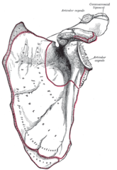
The scapula is a thin, flat triangular-shaped bone placed on the postero-lateral aspect of the thoracic cage. It has 2 surfaces, 3 borders, 3 angles and 3 processes.
Surfaces[edit | edit source]
- Costal Surface or Subscapular Fossa
It is concave and is directed medially and forwards. It is marked by 3 longitudinal ridges. A thick ridge adjoins the lateral border. This part of the bone is almost rod-like acts a lever for the action of serratus anterior in overhead abduction of the arm. - Dorsal Surface
The prominent spine of scapula divides the surface into a smaller supraspinous fossa and a larger infraspinous fossa. The depth of the supraspinous fossa is filled by the supraspinatus muscle.
Borders[edit | edit source]
- Superior Border
It is thin and shorter. It presents the suprascapular notch near the root of the coracoid process. The superior border extends from the superior angle laterally towards coracoid process. - Lateral Border
This is thick and presents infraglenoid tubercle at the upper end. The lateral or axillary border runs from the inferior angle to the lateral angle of the scapula. - Medial Border
This is thin and extends from superior to inferior angle. With the arm resting by the side, the medial or vertebral border runs almost parallel to the spinal column.
Angles[edit | edit source]
- Superior Angle iscovered by trapezius
- Inferior Angle is covered by the latissimus dorsi. It moves forwards round the chest, when the arm is abducted. Palpation of the inferior angle provides a convenient method for following the movement of the scapula during arm motion.
- Lateral or Glenoid Angle is broad and bears the glenoid cavity or fossa, which is directed forward, laterally and slightly upwards.
Processes[edit | edit source]
- Spine or Spinuous Process is a triangular plate of bone with 3 borders and 2 surfaces. It divides the dorsal surface of the scapula into supraspinous and infraspinous fossae. The posterior border is called the crest of the spine which has upper and lower lips.
- The Acromion Process has 2 borders, medial and lateral; 2 surfaces and a facet for clavicle.
- The Coracoid Process


Attachments on Scapula[edit | edit source]
Scapula has various bony projections for attachment of muscles, ligaments and other soft-tissue structures.[2] These include the scapular spine, acromion process, coracoid process, scapular notch, lateral scapular spine, and glenoid fossa. The suprascapular nerve travels through the notch and provides motor supply to the supraspinatus muscle and infraspinatus muscle, as well as sensation to the acromioclavicular joint.[2] [3]
Ossification[edit | edit source]
The scapula ossifies from one primary centre and seven secondary centres. The primary centre appears near the glenoid cavity during the 8th week of development. The 1st secondary centre appears in the middle of coracoid process during the first year and fuses by the 15th year. The subcoracoid centre appears in the root of the coracoid process during the 10th year and fuses by the 16th to 18th years. The other centres, including 2 for the acromion , oe for the lower 2/3rds of the margin of the glenoid cavity, one for the medial border and one for the inferior angle, appear at puberty and fuse by the 25th year. [2]
Soft Tissue[edit | edit source]
Muscles[edit | edit source]
- Subscapularis arises from the medial 2/3rds of the subscapular fossa.
- Supraspinatus arises from medial 2/3rds of supraspinous fossa including upper surface of the spine
- Infraspinatus arises from medial 2/3rds of infraspinous fossa, including lower surface of spinthe e.
- Deltoid arises from lower border of the crest of spinthe e and from lateral border acromion.
- Latissimus Dorsi lower fibres originate from inferior angle of scapthe ula
- Trapezius is inserted into the upper border of the crest of the spine and into medial border of the acromion.
- Serratus anterior is inserted along the medial border of costal surface; 1 digitation from the superior angle to the root of spthe ine, 2 digitations to the medial border, 5 digitations to the inferior angle.
- The long head of biceps brachii arises from supraglenoid tubercle and the short head from the lateral part of the tip of the coracoid process.
- Coracobrachialis arises from medial part of tip ohe tf coracoids process
- Pectoralis minor is inserted into the medial borderand superior surface of coracoidsprocess.
- The long head of triceps arises from infraglenoid tubercle
- Teres minor arises from upper 2/3rds of rough strip on the dorsal surface along the lateral border.
- Teres major arises from lower 1/3rd of rough strip on the dorsal aspect of lateral border
- Levator scapulae is inserted along the dorsal aspect of the medial border, from superior angle up to root of spinthe e
- Rhomboideus minor is inserted into medial border (dorsal aspect) opposite to root of spine
- Rhomboideus major is inserted into the medial border (dorsal aspect) between the root of spine and inferior angle
- Inferior belly of omohyoid arises from upper border near suprthe ascapular notch.
Ligaments[edit | edit source]
- The margin of glenoid cavity gives attachment to the capsule of shouthe lder joint and to the glenoid labrum
- The margin of the facet on the medial aspect of the acromion gives attachment to the capsule of the acromioclavicular joint
- The coracoacromial ligament is attached to the lateral border of the coracoids process and to the medial side of the tip of the acromion process
- The coracohumeral ligament is attached to the root of the coracoids process.
- The coracoclavicular ligament is attached to the coracoid process; the trapezoid part on the superior aspect, and the conoid part near the root. The coracoclavicular ligament is made up of 2 bands: the conoid and the trapezoid, both of which provide vertical stability. The coracoacromial ligament connects the coracoid process to the acromion.
- The suprascapular ligament bridges across the suprascapular notch and converts it into a foramen which transmits the suprascapular nerve. The suprascapular ligament lie above the ligament.
- The spinoglenoid ligayment bridges the spinoglenoid notch. The suprascapular vessels and nerve pass to it.
- The acromioclavicular ligament connects the distal end of the clavicle to the acromion and provides horizontal stability
Bursae[edit | edit source]
There are two major bursae:
- Scapulothoracic Bursa, between the serratus and the thorax, and
- Subscapularis Bursa, between the subscapularis and the serratus.

Biomechanics[edit | edit source]
The scapula upwardly rotates in the frontal plane, posteriorly tilts in the parasagittal plane, and externally rotates in the transverse plane during functional elevation. Scapular control is essential to scapulohumeral coordination. Posterior tilting is responsible for humeral clearance during the acromiohumeral portion of shoulder elevation.
Fung et al discovered that scapular upward rotation and retraction are greatest during abduction elevation, when compared to flexion elevation. They also discovered that posterior tilting was greatest during flexion elevation. Any disturbance in this rhythm can decrease scapulothoracic movement and can be associated with fatigue, impingement, instability, and limits in elevation. [2] [3]
References[edit | edit source]
- ↑ Kenhub-Learn Human Anatomy.Anatomy and Function of the Scapula-Human Anatomy|Kenhub.Available from:https://youtu.be/fEXrPsGySKI (Assessed 31 July 2020)
- ↑ 2.0 2.1 2.2 2.3 Williams GR, Shakil M, Klimkiewicz J. Anatomy of the Scapulothoracic Articulation. Clin Orthop Relat Res. Feb 1999;237-46. [Medline].
- ↑ 3.0 3.1 Culham E, Peat M. Functional Anatomy of the Shoulder Complex. J Orthop Sports Phys Ther. Jul 1993;18(1):342-50. [Medline].

