Femoral Fractures: Difference between revisions
Alex Palmer (talk | contribs) No edit summary |
Kim Jackson (talk | contribs) m (Text replacement - "Femoral stress fracture" to "Femoral stress fracture") |
||
| (64 intermediate revisions by 12 users not shown) | |||
| Line 1: | Line 1: | ||
<div class="editorbox">'''Original Editors ''' - [[User:Vanderpooten Willem|Willem Vanderpooten]] | <div class="editorbox"> '''Original Editors ''' - [[User:Vanderpooten Willem|Willem Vanderpooten]] | ||
'''Top Contributors''' - {{Special:Contributors/{{FULLPAGENAME}}}} | '''Top Contributors''' - [http://www.physio-pedia.com/User:Margaux_Jacobs Margaux Jacobs,] {{Special:Contributors/{{FULLPAGENAME}}}} | ||
</div> | </div> | ||
== | == Introduction == | ||
[[File:Femoral-shaft-fracture.jpeg|thumb|Femoral-shaft-fracture]] | |||
The [[femur]] is the largest and strongest [[bone]] in the body. Due to its strength it requires a significant force to break it. However, certain medical conditions that weaken the bone make it more vulnerable to fracture, so called [[Insufficiency Fracture|pathological fractures]]. For example: [[osteoporosis]]; [[Oncology|malignancy]]; [[Infectious Disease|infection]].<ref>Very well health Femur Fracture Available;https://www.verywellhealth.com/femur-fracture-2549281 (accessed 10.12.2022)</ref> | |||
[[File:Neck of femur fracture (garden IV).jpeg|thumb|Neck of femur fracture]] | |||
== Location == | |||
[[File:Distal-femoral-fracture.png|thumb|299x299px|Distal-femoral-fracture]] | |||
There are different types of femoral fractures according where they occur, namely: | |||
# Femoral Head Fractures: often seen in the elderly osteoporotic population where the cortical bone is weak and so is the trabecular system. It occurs spontaneously or due to low-energy trauma. In the younger population, this fracture is rare and occurs due to high-energy trauma, usually associated with [[Hip Dislocation|hip dislocations]].<ref>Orthobullets Femoral Head Fractures Available:https://www.orthobullets.com/trauma/1036/femoral-head-fractures (accessed 10.12.2022)</ref> | |||
#[[Femoral Neck Hip Fracture|Femoral Neck Fractures]]: one of the most frequent fractures presenting to the emergency department and orthopedic trauma teams.<ref>[[Femoral Neck Hip Fracture|Hip Fracture]]</ref>See link for more. | |||
# [[Femoral Shaft Fractures]]: more common in men after a high-energy impact or in elderly women after a low-energy fall. <ref>Mercer's Textbook of Orthopaedics and Trauma Tenth edition edited by Suresh Sivananthan, Eugene Sherry, Patrick Warnke, Mark D Miller</ref> They can be described as follows: Type I - Spiral or transverse (most common) Type II – Comminuted Type III - Open <ref>WebMD. Broken bone: types of fractures, symptoms and prevention. 2014. (Available at http://www.webmd.boots.com/a-to-z-guides/bone-fractures-types-symptoms-prevention, accessed on 21 december 2014)fckLR</ref> | |||
# [[Distal femoral fracture]]: involve the femoral condyles and the metaphyseal region, often the resulting from high energy trauma eg motor vehicle accidents or a fall from a height. In the elderly, they may occur as an accident at home eg [[Falls in elderly|fall]].<ref>Radiopedia Distal femoral fracture. Available: https://radiopaedia.org/articles/distal-femoral-fracture (accessed 10.12.2022)</ref>See link for more. | |||
== Types == | |||
There are 4 types of fracture: | |||
== | # [[Femoral Stress Fracture|Femoral stress fracture]] | ||
# Severe impaction fractures: the bone breaks into multiple fragments, which are driven into each other. It is a closed fracture that occurs when pressure is applied to both ends of the bone, causing it to split into two fragments that jam into each other. <ref>2.0 2.1 WebMD. Understanding Bone Fractures - the Basics. 2014. (Available at http://www.webmd.com/a-to-z-guides/understanding-fractures-basic-information, accessed 19 December 2014) fckLR</ref> | |||
# Partial fracture: incomplete break of a bone. This type of fracture refers to the way the bone breaks. In an incomplete fracture, the bone cracks but doesn’t break all the way through. In contrast, there is a complete fracture, where the bone snaps into two or more parts.<ref>Cleveland Clinic. Fractures. 2013. (Available at http://my.clevelandclinic.org/services/orthopaedics-rheumatology/diseases-conditions/hic-fractures, assessed 21 December 2014)fckLR</ref><ref>WebMD. Broken bone: types of fractures, symptoms and prevention. 2014. (Available at http://www.webmd.boots.com/a-to-z-guides/bone-fractures-types-symptoms-prevention, accessed on 21 december 2014)fckLR</ref> | |||
# Completed displaced fracture: breaks into two or more pieces and is no longer correctly aligned. Displacement of fractures is defined in terms of the abnormal position of the distal fracture fragment in relation to the proximal bone. <ref>2.0 2.1 WebMD. Understanding Bone Fractures - the Basics. 2014. (Available at http://www.webmd.com/a-to-z-guides/understanding-fractures-basic-information, accessed 19 December 2014) fckLR</ref> | |||
# | |||
# | |||
== References == | == References == | ||
<references / | <references /> | ||
<br> | |||
[[Category:Injury]] | |||
[[Category:Bones]] | |||
[[Category:Musculoskeletal/Orthopaedics|Orthopaedics]] | |||
[[Category:Conditions]] | |||
[[Category:Hip]] | |||
[[Category:Hip - Conditions]] | |||
[[Category:Older People/Geriatrics]] | |||
[[Category:Older People/Geriatrics - Conditions]] | |||
[[Category:Knee]] | |||
[[Category:Knee - Conditions]] | |||
[[Category:Fractures]] | |||
Latest revision as of 10:02, 10 May 2024
Top Contributors - Margaux Jacobs, Kim Jackson, Vanderpooten Willem, Lucinda hampton, Admin, Vidya Acharya, Nupur Smit Shah, Jentel Van De Gucht, Rachael Lowe, Alex Palmer, Daphne Jackson, Margaux Jacobs, Aminat Abolade, Jason Coldwell, WikiSysop, Lauren Lopez, 127.0.0.1 and Evan Thomas
Introduction[edit | edit source]
The femur is the largest and strongest bone in the body. Due to its strength it requires a significant force to break it. However, certain medical conditions that weaken the bone make it more vulnerable to fracture, so called pathological fractures. For example: osteoporosis; malignancy; infection.[1]
Location[edit | edit source]
There are different types of femoral fractures according where they occur, namely:
- Femoral Head Fractures: often seen in the elderly osteoporotic population where the cortical bone is weak and so is the trabecular system. It occurs spontaneously or due to low-energy trauma. In the younger population, this fracture is rare and occurs due to high-energy trauma, usually associated with hip dislocations.[2]
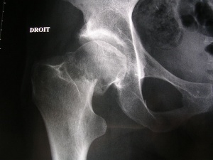
- Femoral Neck Fractures: one of the most frequent fractures presenting to the emergency department and orthopedic trauma teams.[3]See link for more.
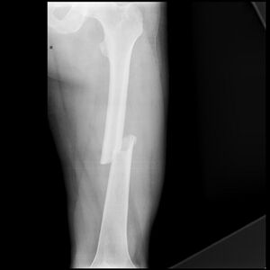
- Femoral Shaft Fractures: more common in men after a high-energy impact or in elderly women after a low-energy fall. [4] They can be described as follows: Type I - Spiral or transverse (most common) Type II – Comminuted Type III - Open [5]
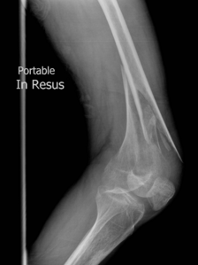
- Distal femoral fracture: involve the femoral condyles and the metaphyseal region, often the resulting from high energy trauma eg motor vehicle accidents or a fall from a height. In the elderly, they may occur as an accident at home eg fall.[6]See link for more.
Types[edit | edit source]
There are 4 types of fracture:
- Femoral stress fracture
- Severe impaction fractures: the bone breaks into multiple fragments, which are driven into each other. It is a closed fracture that occurs when pressure is applied to both ends of the bone, causing it to split into two fragments that jam into each other. [7]
- Partial fracture: incomplete break of a bone. This type of fracture refers to the way the bone breaks. In an incomplete fracture, the bone cracks but doesn’t break all the way through. In contrast, there is a complete fracture, where the bone snaps into two or more parts.[8][9]
- Completed displaced fracture: breaks into two or more pieces and is no longer correctly aligned. Displacement of fractures is defined in terms of the abnormal position of the distal fracture fragment in relation to the proximal bone. [10]
References[edit | edit source]
- ↑ Very well health Femur Fracture Available;https://www.verywellhealth.com/femur-fracture-2549281 (accessed 10.12.2022)
- ↑ Orthobullets Femoral Head Fractures Available:https://www.orthobullets.com/trauma/1036/femoral-head-fractures (accessed 10.12.2022)
- ↑ Hip Fracture
- ↑ Mercer's Textbook of Orthopaedics and Trauma Tenth edition edited by Suresh Sivananthan, Eugene Sherry, Patrick Warnke, Mark D Miller
- ↑ WebMD. Broken bone: types of fractures, symptoms and prevention. 2014. (Available at http://www.webmd.boots.com/a-to-z-guides/bone-fractures-types-symptoms-prevention, accessed on 21 december 2014)fckLR
- ↑ Radiopedia Distal femoral fracture. Available: https://radiopaedia.org/articles/distal-femoral-fracture (accessed 10.12.2022)
- ↑ 2.0 2.1 WebMD. Understanding Bone Fractures - the Basics. 2014. (Available at http://www.webmd.com/a-to-z-guides/understanding-fractures-basic-information, accessed 19 December 2014) fckLR
- ↑ Cleveland Clinic. Fractures. 2013. (Available at http://my.clevelandclinic.org/services/orthopaedics-rheumatology/diseases-conditions/hic-fractures, assessed 21 December 2014)fckLR
- ↑ WebMD. Broken bone: types of fractures, symptoms and prevention. 2014. (Available at http://www.webmd.boots.com/a-to-z-guides/bone-fractures-types-symptoms-prevention, accessed on 21 december 2014)fckLR
- ↑ 2.0 2.1 WebMD. Understanding Bone Fractures - the Basics. 2014. (Available at http://www.webmd.com/a-to-z-guides/understanding-fractures-basic-information, accessed 19 December 2014) fckLR


