Achilles Tendinopathy: Difference between revisions
Eva Roose1 (talk | contribs) No edit summary |
Eva Roose1 (talk | contribs) No edit summary |
||
| Line 46: | Line 46: | ||
<br>So we can say that the effects of overuse, poor circulation, lack of flexibility, gender, endocrine or metabolic factors can lead to tendinopathies. The structure of the tendon is disturbed by this repetitive strain (often eccentric nature) and collagen fibers go along together, break the crosslinks and the slide denaturation cause tissue, causing inflammation. This cumulative microtrauma is assumed not only weaken collagen cross-linking, but also the collagenous matrix and the vascular elements of influencing the tendon, which ultimately leads to tendonitis.[16]<br><span style="font-size: 13px;" /> | <br>So we can say that the effects of overuse, poor circulation, lack of flexibility, gender, endocrine or metabolic factors can lead to tendinopathies. The structure of the tendon is disturbed by this repetitive strain (often eccentric nature) and collagen fibers go along together, break the crosslinks and the slide denaturation cause tissue, causing inflammation. This cumulative microtrauma is assumed not only weaken collagen cross-linking, but also the collagenous matrix and the vascular elements of influencing the tendon, which ultimately leads to tendonitis.[16]<br><span style="font-size: 13px;" /> | ||
== | == Clinically Relevant Anatomy<br> == | ||
[[Image:Achilles tendon.jpg|thumb|right|200px|Achilles Tendon]] | [[Image:Achilles tendon.jpg|thumb|right|200px|Achilles Tendon]] | ||
The | The achilles tendon is the biggest and strongest tendon in the human body. The tendon has the capacity to resist large tensile forces. It stems from a distal confluence of the gastrocnemius and soleus muscle and inserts at the bottom of the calcaneus. Like other tendons, Achilles has a hierarchical structure. [12] | ||
The M. soleus is a prime mover plantar flexor of the ankle and the m. gastrocnemius is a flexor of the knee. | |||
A typical tendon structure consists of thin, cylindrical cells and an extracellular matrix. The cells of the tendon, respectively tenocytes tenoblasts, are responsible for the synthesis of all of the components of the extracellular matrix. Inside the matrix we find bundles of type I collagen and elastin. This type-I collagen is responsible for the strength of the tendon. Between the collagen there is a ground substance located which is made up of proteoglycans and glycosaminoglycans. [13] | |||
The achilles tendon is surrounded by parthenon and not by the common known synovial membrane. The parthenon works as an elastic sleeve around the tendon which allows the tendon to move freely between surrounding tissue. The parthenon consists out a layer of cells and is responsible for blood transportation of the tendon. The layers that are formed under the parthenon are in chronological order: the epitenon which is a thin membrane and the endotenon wichon his turn surrounds the collagen fibers which results in bundles. [14]<br> | |||
== Mechanism of Injury / Pathological Process<br> == | == Mechanism of Injury / Pathological Process<br> == | ||
Revision as of 20:07, 22 December 2016
Original Editor - Karolyn Conolty
Top Contributors - Aline Tréfois, Admin, Puja Gaikwad, Kim Jackson, Eva Roose1, Rachael Lowe, Lucinda hampton, Laura Ritchie, Shauni Van Overstraeten, Karolyn Conolty, Chi Ngai LO, Christopher, Fasuba Ayobami, Simisola Ajeyalemi, Birgit Schauvliege, Wanda van Niekerk, Naomi O'Reilly, 127.0.0.1, Vidya Acharya, Rucha Gadgil, Eric Robertson, Samuel Adedigba, Jess Bell, Khloud Shreif, David Bayard, Camille Linussio and Olajumoke Ogunleye
Search Strategy [edit | edit source]
We began with the template of the VUB. We searched articles based on the 16 contents. Some contents already had information about our disease on the current Physiopedia page and other contents didn’t. We added the information from the articles to the existing text.
Databases searched: VUBis, Pubmed, Physiopedia, JOSPT, Google Scholar, British Journal of Sport Medicine, Web of Science, Fai sagepub
Keywords searched:
achilles tendonitis
achilles tendinopathy
achilles tendinitis
clinical characteristics and achilles tendonitis
achilles tendonitis treatment
achilles tendonitis physical therapy
physiotherapy and achilles tendonitis
differential diagnose and achilles tendinopathy
achilles tendinopathy and diagnostic procedures
...
Definition/Description [edit | edit source]
Achilles tendinitis is a common overuse injury caused by an excessive stretching or tearing of the Achilles tendon in an acute context. This can lead to a sudden injury, or in the worst case, can cause a rupture of the Achilles tendon. In both cases, a lack of flexibility or a stiff Achilles tendon can increase the risk of these injuries. [1]
A lot of descriptions are given concerning Achilles Tendinopathy. Usually a difference is made between the insertional and the mid portion form. The difference is in the localisation. The insertional form is situated at the level of transition between the Achilles tendon and the bone. The midportion form is located at the level of the tendon body. It is described as the gradually emerged of pain 2-6 cm above the calcaneal insertion of the Achilles tendon, associated with degenerative changes and often with thickening of the tendon (Alfredson, 2003; Jozsa et al., 1997; Movin et al., 1997; Paavola et al., 2002a). [2]
Surgical specimens show a range of degenerative changes of the affected tendon, such as in the tendon fibre structure and arrangement as well as an increase in glycosaminoglycans, which may explain the swelling of the tendon. [3] The precise cause of tendonitis remains unclear. Even though tendonitis of the achilles tendon is often connected to sport activities, the ailment is also often found with people who do not practice sports. The biggest cause is the excessive overburdening of the tendon. A light degeneration of the achilles tendon can be latently present, but pain only appears when the tendon is overburdened. It is also noted that the ailment is usually not preceded by a trauma. [4], [5]
The terms “tendinose”, “tendonitis” or “tendinitis” and “tendinopathy” are commonly used. (Paavola et al., 2002a). The definitions of those terms are often described in a different manner. This makes it difficult to evaluate articles and to compare them (Alfredson, 2003). [6]
Tendinitis is the inflammation of the tendon and is caused by micro-tears that happen when the musculotendinous unit is acutely overloaded with a tensile force that is too heavy and/or too sudden. [7]
Achilles tendonitis is commonly seen in athletes who sustain an increase in training load, and is most often due to overuse. Tendons respond poorly to overuse, therefore healing is slow. This can leave a tendon pathologically defective, which decreases tendon strength and leaves it less able to tolerate load, thus vulnerable to further injury [8] or tendinosis. Extrinsic factors contributing to this condition include training errors and inappropriate footwear. Intrinsic factors include inflexibility, weakness and malalignment. [9]
In other situations, there will be clinical inflammation, but objective pathologic evidence for cellular inflammation is lacking, and in these conditions the term tendonitis is more appropriate. Tendonitis is a degeneration of the tendon’s collagen in response to chronic overuse; when overuse is continued without giving the tendon time to heal and rest, such as with repetitive strain injury. Even tiny movements, such as clicking on a mouse, can cause tendinosis, when done repeatedly.
Achilles tendonitis is a degenerative change of the Achilles Tendon associated with pain and often with the thickening of the tendon. It is common in athletes, but it also occurs in non athletes. Surgical specimens show a range of degenerative changes of the affected tendon, such as changes in tendon fibre structure and arrangement as well as an increase in glycosaminoglycans, which may explain the swelling of the tendon. [3] The precise cause of tendonitis remains unclear. Even though tendonitis of the achilles tendon is often connected to sport activities, the ailment is also often found with people who do not practice sports. The biggest cause is the excessive overburdening of the tendon. A light degeneration of the achilles tendon can be latently present, but pain only comes into being when the tendon is overburdened. It is also noted that the ailment is usually not preceded by a trauma. [4], [5]
Tendonitis is often confused with tendonitis, but it is important to understand the difference between these two pathologies. Tendonitis is an inflammation of the tendon. This inflammation causes micro-tears in the tendon when it is acutely overloaded. This diagnosis is often mistakenly used when the patient actually has tendonitis. Tendonitis is a degeneration process in which no temperature rises occur, as opposed to tendonitis. It is very important to distinguish between these disorders, to discover which treatment is required, and what the expected duration of the treatment will be.
The healing time for tendonitis is generally shorter, and commonly takes several days to 6 weeks. For tendinosis, the expected duration is variable, this can be 6-10 weeks, but it might also take 3-6 months, when the tendonitis has become chronic. [10], [11]
Epidemiology /Etiology [edit | edit source]
The etiology of Achilles tendonitis is still unclear. Straining the tendon during physical exercise has been seen as one of the biggest pathological stimulus. Systematic overloading of the Achilles tendon above his physiological limit can cause a micro-trauma.
Repetitive micro-traumas that are linked with a non-uniform tension between the M. gastrocnemius and M.soleus, cause frictional forces between the fibers and abnormal concentrations of the loading in the achilles tendon. This has consequences such as the inflammation of the tendon sheath, degeneration, or a combination of both. Without the minimum time for recovery, this can lead to a tendinopathy. [15]
In acute trauma, the external factors dominate , while injuries caused by overuse generally have a multifactorial origin. The acute phase of Achilles tendinopathy is caused by acute overload, blunt trauma or acute muscle fatigue, and is characterized by an inflammatory reaction and edema formation. If the treatment of the acute phase fails or if they overlooked it, it can cause a fibrin and form adhesions off the tendon.
Often, tendon degeneration is found in combination with peritendinous adhesions, but this does not mean that one condition causes the other one. Decreased arterial blood flow, local hypoxia, decreased metabolic activity, nutrition, and a persistent inflammatory response have been suggested as possible factors that could lead to chronic tendon overuse injuries and tendon degeneration.
In epidemiological studies, various alignments and biomechanical faults are claimed to play a causative role in two-thirds of the athletes with an Achilles tendon disorder.
The most common and perhaps the most important malalignment is the one of the ankle caused by hyperpronation of the foot. Increased foot pronation has been proposed to be associated with Achilles tendinopathy.
So we can say that the effects of overuse, poor circulation, lack of flexibility, gender, endocrine or metabolic factors can lead to tendinopathies. The structure of the tendon is disturbed by this repetitive strain (often eccentric nature) and collagen fibers go along together, break the crosslinks and the slide denaturation cause tissue, causing inflammation. This cumulative microtrauma is assumed not only weaken collagen cross-linking, but also the collagenous matrix and the vascular elements of influencing the tendon, which ultimately leads to tendonitis.[16]
Clinically Relevant Anatomy
[edit | edit source]

The achilles tendon is the biggest and strongest tendon in the human body. The tendon has the capacity to resist large tensile forces. It stems from a distal confluence of the gastrocnemius and soleus muscle and inserts at the bottom of the calcaneus. Like other tendons, Achilles has a hierarchical structure. [12]
The M. soleus is a prime mover plantar flexor of the ankle and the m. gastrocnemius is a flexor of the knee.
A typical tendon structure consists of thin, cylindrical cells and an extracellular matrix. The cells of the tendon, respectively tenocytes tenoblasts, are responsible for the synthesis of all of the components of the extracellular matrix. Inside the matrix we find bundles of type I collagen and elastin. This type-I collagen is responsible for the strength of the tendon. Between the collagen there is a ground substance located which is made up of proteoglycans and glycosaminoglycans. [13]
The achilles tendon is surrounded by parthenon and not by the common known synovial membrane. The parthenon works as an elastic sleeve around the tendon which allows the tendon to move freely between surrounding tissue. The parthenon consists out a layer of cells and is responsible for blood transportation of the tendon. The layers that are formed under the parthenon are in chronological order: the epitenon which is a thin membrane and the endotenon wichon his turn surrounds the collagen fibers which results in bundles. [14]
Mechanism of Injury / Pathological Process
[edit | edit source]
Tendonitis[edit | edit source]
Tendinitis is the inflammation of the tendon and results from micro-tears that happen when the musculotendinous unit is acutely overloaded with a tensile force that is too heavy and/or too sudden[1].
Achilles tendonitis is commonly seen in athletes who sustain an increase in training load, and is most often due to overuse. Tendons respond poorly to overuse, therefore healing is slow. This can leave a tendon pathologically defective, which decreases tendon strength and leaves it less able to tolerate load, thus vulnerable to further injury[2] or tendinosis. Extrinsic factors contributing to this condition include training errors and inappropriate footwear. Intrinsic factors include inflexibility, weakness and malalignment. [3]
Tendinosis[edit | edit source]
In other situations, there will be clinical inflammation, but objective pathologic evidence for cellular inflammation is lacking, and in these conditions the term tendinosis is more appropriate. Tendinosis is a degeneration of the tendon’s collagen in response to chronic overuse; when overuse is continued without giving the tendon time to heal and rest, such as with repetitive strain injury, tendinosis results. Even tiny movements, such as clicking a mouse, can cause tendinosis, when done repeatedly.
Achilles tendinosis is a degenerative change of the Achilles Tendon associated with pain and often with thickening of the tendon. It is common in athletes, but it also occurs in non athletes. Surgical specimens show a range of degenerative changes of the affected tendon, such as changes in tendon fibre structure and arrangement as well as an increase in glycosaminoglycans, which may explain the swelling of the tendon.[4] The precise cause of tendinosis remains unclear. Even though tendinosis of the achilles tendon is often connected to sport activities, the ailment is also often found with people who do not practice sports. The biggest cause is the excessive overburdening of the tendon. A light degeneration of the achilles tendon can be latently present, but pain only comes into being when the tendon is overburdened. It is also noted that the ailment is usually not preceded by a trauma.[5], [6]
Tendinosis is often confused with tendonitis, but it is important to understand the difference between these two pathologies. Tendonitis is an inflammation of the tendon. This inflammation causes micro-tears in the tendon when the tendon is acutely overloaded. This diagnosis is often mistakenly used when the patiënt actually has tendinosis. Tendinosis is a degeneration process in which no temperature rises occur, as apposed to tendonitis. It is very important to distinguish between these disorders, to discover which treatment is required, and what the expected duration of the treatment will be.
The healing time for tendonitis is generally shorter, and commonly takes several days to 6 weeks. For tendinosis, the expected duration is variable, this can be 6-10 weeks, but it might also take 3-6 months, when the tendinosis has become chronic. [7], [8]
Characteristics/Clinical Presentation[edit | edit source]
Morning pain is a hallmark symptom because the achilles tendon must tolerate full range of movement including stretch immediately on rising in the morning. Symptoms are typically localized to the tendon and immediate surrounding area. Swelling and pain at the attachment are less common. The tendon can appear to have subtle changes in outline, becoming thicker in the A-P and M-L planes.[2]
With people who have a tendinopathy of the achilles tendon that has a sensitive zone, combined with intratendinous swelling, that moves along with the tendon and of which sensitivity increases or decreases when the tendon is put under pressure, there will be a high predictive value that in this situation there is a case of tendinosis.[9]
Differential Diagnosis
[edit | edit source]
Posterior Ankle Impingement, Medial Tendinopathy, Retrocalcaneal Bursitis, Sural Nerve, Lumbar Radiculopathy, Ankle OA, DVT, Haglund Deformity, Partial Achilles Tendon Rupture.[2]
Diagnostic Procedures[edit | edit source]
Examination of the achilles tendon is inspection for muscle atrophy, swelling, asymmetry, joint effusions and erythema. Atrophy is an important clue to the duration of the tendinopathy and it is often present with chronic conditions. Swelling, asymmetry and erythema in pathologic tendons are often observed in the examination. Joint effusions are uncommon with tendinopathy and suggest the possibility of intra-articular pathology.
Range of motion testing, strength and flexibility are often limited on the side of the tendinopathy.[10], [11]
Palpation tends to elicit well-localized tenderness that is similar in quality and location to the pain experienced during activity.[12]
Physical examinations of the Achilles tendon often reveals palpable nodules and thickening. Anatomic deformities, such as forefoot and heel varus and excessive pes planus or foot pronation, should receive special attention. These anatomic deformities are often associated with this problem.[13], [14]
In case extra research is wanted, an echography is the first choice of examination when there is a suspicion of tendinosis.[15]
Imaging studies are not necessary to diagnose achilles tendonitis, but may be useful with differential diagnosis. Ultrasound is the imaging modality of first choice as it provides a clear indication of tendon width, changes of water content within the tendon and collagen integrity, as well as bursal swelling. MRI may be indicated if diagnosis is unclear or symptoms are atypical. MRI may show increased signal within the Achilles.[2]
Outcome Measures[edit | edit source]
Robinson et al recommend the VISA-A scale. This is a subjective rating scale that quantifies the symptoms and dysfunction in the Achilles tendon. It is very useful to rate Achilles tendons and to assess progress of recovery during rehabilitation. [2][16]
Examination[edit | edit source]
add text here related to physical examination and assessment
Medical Management
[edit | edit source]
add text here
Physical Therapy Management / Interventions
[edit | edit source]
The treatment should be conservative, including rest, equipment changes, strength and flexibility exercises. A popular and effective option is the eccentric strength training. Deep friction massage and stretching of the gastrocnemius and soleus are considered helpful for Achilles tedinopathy.[17] Anatomic deformities can be treated with shoe orthotics. These shoe orthotics correct overpronation or pes planus problems.[18]
The effects of physical therapy on achilles tendonitis is poorly understood, although musculotendinous strengthening appears essential. Eccentric exercises have been shown to have positive effects of Achilles tendonitis, and remains the gold standard for rehabiliation of this condition.[2][3] A study by Roos et al concluded that eccentric exercises improve function and reduce pain and effects were apparent after 6 weeks of treatment, lasting for 1 year.[16]
An inflammation is necessary to start a restoration process in the damaged tissue, but the use of certain medication, such as corticosteroids and quinolones counter the inflammation, and as a result also the restoration process. Even when the patient does not take this medication, tendinosis is also a consequence of a disrupted restoration process.[19]
Supportive taping can also help manage symptoms:
| [20] | [21] |
Conservative Treatment: [22],[23][edit | edit source]
In order to treat the symptoms, antiflogistics or other anti-inflammatory therapy are often used. However these forms of therapy usually cannot prevent the injury to live on.
Nevertheless patients will always have to be encouraged to execute less burdening activities, so that the burden on the tendon decreases as well. Complete immobilisation should however be avoided, since it can cause atrophy.
Passive Rehabilitation:
- Mobilisations can be used for dorsiflexion limitation of the talocrural joint and varus- or valgus limitation of the subtalar joint.
- Deep cross frictions (15 min). It’s effectiveness is not scientifically proven and gives limited results. [24], [25], [26], [27]
- Recently, the use of Extracorporal Shock Wave Therapy was proven.[28], [29], [30]
- Besides that, the application of ice can cause a short decrease in pain and in swelling. Even though cryotherapy 2, 5 was not studied very thoroughly, recent research has shown that for injuries of soft tissue, applications of ice through a wet towel for ten minutes are the most effective measures. [31], [32], [33]
Active Rehabilitation:
- An active exercise program mostly includes eccentric exercises. This can be explained by the fact that eccentric muscle training will lengthen the muscle fibres, which stimulates the collagen production. This form of therapy appears successful for mid-portion tendinosis, but has less effect with insertion tendinopathy. The sensation of pain sets the beginning burdening of the patient and the progression of the exercises.[34], [35], [36]
Thorough management guidelines for achilles tendinopathy is covered in detail in the Achilles Tendinopathy Toolkit.
Key Research[edit | edit source]
add links and reviews of high quality evidence here (case studies should be added on new pages using the case study template)
Clinical Bottom Line[edit | edit source]
add text here
Resources[edit | edit source]
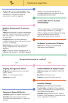 |
Achilles Tendinopathy Toolkit
The Achilles Tendinopathy Toolkit is a comprehensive evidence based resource to assist practitioners in clinical decision making for Achilles Tendinopathy. |
Recent Related Research (from Pubmed)[edit | edit source]
Failed to load RSS feed from http://eutils.ncbi.nlm.nih.gov/entrez/eutils/erss.cgi?rss_guid=1DeUIcPgNLDEQT17PEJLA26uuKLv4fz4eTsAi9nkGXzfWOXsZF|charset=UTF-8|short|max=10: Error parsing XML for RSS
Presentations[edit | edit source]
 |
Achilles Tendonopathy: Intervention
This presentation, created by Shannon Petersen, Clebert LeBlanc, Amy Lavrich, & Kelly Coleman as part of the Regis University OMPT Fellowship, discusses the current best evidence for interventions for Achilles Tendonopathy. |
Read 4 Credit[edit | edit source]
 |
Would you like to earn certification to prove your knowledge on this topic? All you need to do is pass the quiz relating to this page in the Physiopedia member area.
|
References[edit | edit source]
References will automatically be added here, see adding references tutorial.
- ↑ Evelyn Bass. Tendinopathy: Why the Difference Between Tendinitis and Tendinosis Matters Int J Ther Massage Bodywork. 2012; 5(1): 14–17.fckLRPublished online Mar 31, 2012.
- ↑ 2.0 2.1 2.2 2.3 2.4 2.5 Cite error: Invalid
<ref>tag; no text was provided for refs namedCook et al - ↑ 3.0 3.1 Roos EM, Engstrom M, Lagerquist A, Soderberg B. Clinical improvement after 6 weeks of eccentric exercise in patients with mid-portion Achilles tendinopathy - a randomized trial with 1-year follow-up. Scand J Med Sci Sports. 2004;14:286-295.
- ↑ T E O Schubert, C. W. (2005). Achilles tendinosis is associated with sprouting of substance P positive nerve fibres. Ann Rheum Dis , 1083-1086.fckLR Level 3B
- ↑ John J. Wilson, T. M. (2005). Common overuse tendon problems: A review and recommendations for treatment. University of Wisconsin Medical School, Madison, Wisconsin , 1-8.fckLR Level 1A
- ↑ MIKA PAAVOLA, M. P. (2002). Current Concepts Review Achilles Tendinopathy . THE JOURNAL OF BONE AND JOINT SURGERY, INCORPORATED , 2062-2076.
- ↑ K M Khan, a. p. (2002). Time to abandon the “tendinitis” myth: Painful, overuse tendon conditions have a non-inflammatory pathology . BMJ , 324:626.fckLR Level 2A
- ↑ Evelyn Bass, L. (2012). Tendinopathy: Why the Difference Between Tendinitis and Tendinosis Matters. Int J Ther Massage Bodywork. , 5(1): 14–17.fckLR Level 2C
- ↑ KL. Luscombe, P. S. (2003). Achilles tendinopathy. Trauma , 215-225.fckLR Level 2C
- ↑ . Hammer, W. I. (1999). Functional Soft Tissue Examination and Treatment by Manual Methods. Aspen: Jones &amp;amp;amp;amp;amp;amp;amp;amp;amp;amp;amp;amp;amp;amp;amp;amp; Bartlett Learning.
- ↑ KL. Luscombe, P. S. (2003). Achilles tendinopathy. Trauma , 215-225.fckLR Level 2C
- ↑ John J. Wilson, T. M. (2005). Common overuse tendon problems: A review and recommendations for treatment. University of Wisconsin Medical School, Madison, Wisconsin , 1-8.fckLR Level 1A
- ↑ John J. Wilson, T. M. (2005). Common overuse tendon problems: A review and recommendations for treatment. University of Wisconsin Medical School, Madison, Wisconsin , 1-8.fckLR Level 1A
- ↑ Shibuya N, T. J. (2012). Is calcaneal inclination higher in patiënts with insertional achilles tendinosis? A case- controlled, cross-sectional study. The journal of foot and ankle surgery , 757-761.fckLR Level 3B
- ↑ Healy, N. T. (2010). Ultrasound-guided treatments for chronic Achilles tendinopathy: an update and current status . Skeletal Radiol , 39:425–434.fckLR Level 5
- ↑ 16.0 16.1 Robinson JM, Cook JL, Purdam C et al. The VISA-A questionnaire: a valid and reliable index of the clinical severity of Achilles tendinopathy. British J of Sports Med. 2001;35:335-341.
- ↑ John J. Wilson, T. M. (2005). Common overuse tendon problems: A review and recommendations for treatment. University of Wisconsin Medical School, Madison, Wisconsin , 1-8.fckLR Level 1A
- ↑ John J. Wilson, T. M. (2005). Common overuse tendon problems: A review and recommendations for treatment. University of Wisconsin Medical School, Madison, Wisconsin , 1-8.fckLR Level 1A
- ↑ MIKA PAAVOLA, M. P. (2002). Current Concepts Review Achilles Tendinopathy . THE JOURNAL OF BONE AND JOINT SURGERY, INCORPORATED , 2062-2076.
- ↑ Jenna Beaudry. Achilles Tendonitis Tape Job. Available from: http://www.youtube.com/watch?v=xzRhIyw85Xk [last accessed 01/12/12]
- ↑ Aaron Tomlinson. Achilles Tape Application. Available from: http://www.youtube.com/watch?v=fQAwpCToR48 [last accessed 01/12/12]
- ↑ Alex Scott, R. P. (2011). Conservative treatment of chronic Achilles tendinopathy. CMAJ , 183(10): 1159–1165.fckLR Level 1A
- ↑ John J. Wilson, T. M. (2005). Common overuse tendon problems: A review and recommendations for treatment. University of Wisconsin Medical School, Madison, Wisconsin , 1-8.fckLR Level 1A
- ↑ . Hammer, W. I. (1999). Functional Soft Tissue Examination and Treatment by Manual Methods. Aspen: Jones &amp;amp;amp;amp;amp;amp;amp;amp;amp;amp;amp;amp;amp;amp;amp;amp;amp; Bartlett Learning.
- ↑ Stasinopoulos D, S. I. (2004). Comparison of effects of exercise programme, pulsed ultrasound and transverse friction in the treatment of chronic patellar tendinopathy. Clin Rehabil , 18(4):347-52.fckLR Level 1B
- ↑ James Henry Cyriax, P. J. (1993). Illustrated manual of orthopaedic medicine. Oxford: Elsevier Health Sciences.
- ↑ Joseph MF, T. K. (2012). Deep friction massage to treat tendinopathy: a systematic review of a classic treatment in the face of a new paradigm of understanding. J Sport Rehabil. , 21(4):343-53.fckLR Level 3A
- ↑ John J. Wilson, T. M. (2005). Common overuse tendon problems: A review and recommendations for treatment. University of Wisconsin Medical School, Madison, Wisconsin , 1-8.fckLR Level 1A
- ↑ Rompe JD, F. J. (2008). Eccentric loading compared with shock wave treatment for chronic insertional achilles tendinopathy. A randomized, controlled trial. J Bone Joint Surg Am. , (1):52-61.fckLR Level 1B
- ↑ Sten Rasmussen, M. C. (2008). Shockwave therapy for achilles tendinopathy. A double-blind, randomized clinical trail of efficacy. Acta Orthopaedica , 249-256.fckLR Level 1B
- ↑ Bleakley C, M. S. (2004). The use of ice in the treatment of acute soft-tissue injury: a systematic review of randomized controlled trials. Am J Sports Med , (1):251-61.fckLR Level 1A
- ↑ Dykstra JH, H. H. (2009). Comparisons of cubed ice, crushed ice, and wetted ice on intramuscular and surface temperature changes. J Athl Train. , (2):136-41.fckLR Level 2A
- ↑ John J. Wilson, T. M. (2005). Common overuse tendon problems: A review and recommendations for treatment. University of Wisconsin Medical School, Madison, Wisconsin , 1-8.fckLR Level 1A
- ↑ Bleakley C, M. S. (2004). The use of ice in the treatment of acute soft-tissue injury: a systematic review of randomized controlled trials. Am J Sports Med , (1):251-61.fckLR Level 1A
- ↑ Brett L Woodley, R. J.‐W. (2007). Chronic tendinopathy: effectiveness of eccentric exercise. Br J Sports Med , 41(4): 188–198.fckLR Level 1A
- ↑ John J. Wilson, T. M. (2005). Common overuse tendon problems: A review and recommendations for treatment. University of Wisconsin Medical School, Madison, Wisconsin , 1-8.fckLR Level 1A

