Bone
Original Editors -
Top Contributors - Lucinda hampton, Candace Goh, Admin, Shaimaa Eldib, Kim Jackson, Wataru Okuyama, Jess Bell, Robin Tacchetti, George Prudden, Claire Knott, Manisha Shrestha, Khloud Shreif, Sai Kripa, Tony Lowe, Stephanie Geeurickx, WikiSysop and Joao Costa
Introduction[edit | edit source]
The adult human skeleton is composed of 206 bones. At birth, there are approximately 270 bones, with the final adult count decreasing as a portion of these bones fuse during phases of skeletal growth and maturation.
- Bone is a metabolically active connective tissue that provides structural support, facilitates movement, and protects vital organs.
- It plays an important role in regulating mineral and acid-base balance homeostasis. It also provides the environment for hematopoiesis (blood cells production) within the bone marrow.
- Bone is composed of an extracellular matrix and bone cells (osteocytes)[1]. Under the right conditions, bone tissue undergoes a process of mineralization, formed by collagen matrix and hardened by deposited calcium. Bone tissue (osseous tissue) differs greatly from other tissues in the body. Bone is hard and many of its functions depend on that characteristic hardness.[2]The skeleton is composed of around 80% cortical bone and 20% trabecular bone.[3]
- Much of the cellular activity in a bone consists of removal and replacement at the same site, a process called remodelling. The remodelling process occurs throughout life and becomes dominant by the time that bone reaches its peak mass (typically by the early 20s). Remodelling continues throughout life so that most of the adult skeleton is replaced about every 10 years.[4]
Structure[edit | edit source]
The four general categories of bones are:
- long bones : include the clavicles, humeri, radii, ulnae, metacarpals, femurs, tibiae, fibulae, metatarsals, and phalanges
- short bones: include the carpal and tarsal bones, patellae, and sesamoid bones.
- flat bones: include the skull, mandible, scapulae, sternum, and ribs.
- irregular bones. include the vertebrae, sacrum, coccyx, and hyoid bone.
Flat bones form by membranous bone formation, whereas long bones are formed by a combination of endochondral and membranous bone formation.[5]
Gross anatomy:[edit | edit source]

A long bone has two parts: the diaphysis and the epiphysis. The diaphysis is the tubular shaft that runs between the proximal and distal ends of the bone. The hollow region in the diaphysis is called the medullary cavity, which is filled with yellow marrow. The walls of the diaphysis are composed of dense and hard compact bone.[6]
Individual bone structure[edit | edit source]
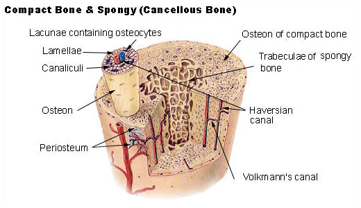
Two types of bone tissue are present - Compact/cortical bone and cancellous/spongy bone.
- Compact/cortical bone - Makes up outer part of bone. Rigid, dense, highly organised bone tissue, arranged in haversian systems which are microscopic cylinders of bone matrix with osteocytes in concentric rings around central haversian canals.
- Cancellous/spongy bone - Makes up inner part of bone. More elastic and porous, storage of red bone marrow. Osteocytes, matrix and blood vessels are not arranged in haversian systems.

Cellular structure;[edit | edit source]
- Osteoblasts - produce matrix (osteoid), build up bone tissue.
- Osteoclasts - secretes acids and enzymes to breakdown bone tissue.
- Osteocytes - maintain bone tissue by mineralising osteoid.[7]
Molecular structure:[edit | edit source]
Approximately 20% of in vivo bone is water. Of the dry bone mass, 60-70% of is bone mineral in the form of small crystals and the rest is collagen. The composition of the mineral component is hydroxyapatite Ca10(PO4)6(OH)2 and collagen is the main fibrous protein of the human body.
Functions:[edit | edit source]
Mechanical :[8][edit | edit source]
- Protect internal organs
- Give shape and support to the body
- Movement
Synthetic:[edit | edit source]
- Manufactures blood cells from the bone marrow (haematopoiesis)
Metabolic;[5][edit | edit source]
- Mineral storage
- Fat storage
- Role in acid-base balance
Remodelling:[edit | edit source]
Purpose:[edit | edit source]
- To allow bone to ordinarily adjust strength in proportion to the degree of bone stress. When subjected to heavy loads, bones will consequently thicken.
- To rearrange shape of bone for proper support of mechanical forces in accordance with stress patterns.
- To replace old bone which may be brittle/weak in order to maintain toughness of bone. New organic matrix is needed as the old organic matrix degenerates[9].
Calcium homeostasis/balance must exist between osteoclasts and osteoblasts activity[edit | edit source]
- If too much new tissue is formed, the bones become abnormally large and thick (acromegaly)
- Excessive loss of calcium weakens the bones, as occurs in osteoporosis
Repair:[edit | edit source]
- The osteoclasts function to remove fragments of dead or damaged bone by dissolving and reabsorbing calcium salts of bone matrix. Like a building that has just collapsed, the rubble must be removed before reconstruction can take place. Osteoblasts are activated to knit the broken ends of bone together.[5]
Clinical Significance[edit | edit source]
Bone tissue is susceptible to a myriad of pathologies that may range from etiologies of embryological, metabolic, autoimmune, neoplastic, or idiopathic origins. These include, but are not limited to, the conditions discussed below.
- Achondroplasia is a genetic disorder commonly associated as a cause of dwarfism. Individuals affected may present with short extremities due to decreased development of endochondral bone.
- Paget disease of the bone is characterized to be an imbalance amongst the activities of osteoblasts and osteoclasts. Of unknown etiology, the condition only affects localized portions of the skeletal tissue, generally involving one or more neighboring bones rather than the diffuse skeletal system.
- If untreated, Paget disease of the bone can act as a risk factor for osteosarcoma, a malignant proliferation of osteoblasts.
- Skeletal neoplasm begins in the metaphysis of long bones with patients complaining of bone pain with swelling or as a pathologic fracture (a break in the bone caused by weakness of the bone through disease rather than trauma). Much more common in teenagers than in the elderly.[10]
- Bone fractures
- Osteoporosis
- Osteoarthritis
- Osteomalacia
- Rickets
- Epiphyseal plate disorders
References[edit | edit source]
- ↑ El Sayed SA, Nezwek TA, Varacallo M. Physiology, Bone. InStatPearls [Internet] 2019 Jul 29. StatPearls Publishing. Available from:https://www.ncbi.nlm.nih.gov/books/NBK441968/ (last accessed 10.2.2020)
- ↑ Opentextbc.ca. (2018). 6.3 Bone Structure – Anatomy and Physiology. [online] Available at: https://opentextbc.ca/anatomyandphysiology/chapter/6-3-bone-structure/
- ↑ Singh S, Bray TJ, Hall-Craggs MA. Quantifying bone structure, micro-architecture, and pathophysiology with MRI. Clinical radiology. 2018 Mar 1;73(3):221-30.
- ↑ Office of the Surgeon General (US. The Basics of Bone in Health and Disease. InBone Health and Osteoporosis: A Report of the Surgeon General 2004. Office of the Surgeon General (US). Available from:https://www.ncbi.nlm.nih.gov/books/NBK45504/ (last accessed 10.2.2020)
- ↑ 5.0 5.1 5.2 Clarke B. Normal bone anatomy and physiology. Clinical journal of the American Society of Nephrology. 2008 Nov 1;3(Supplement 3):S131-9.
- ↑ Hall JE. Guyton and Hall textbook of medical physiology e-Book. Elsevier Health Sciences; 2015 May 31.
- ↑ Burr DB. Targeted and nontargeted remodeling. Bone. 2002;30:2-4.
- ↑ Mackiewicz Z, Niklińska WE, Kowalewska J, Chyczewski L. Bone as a source of organism vitality and regeneration. Folia histochemica et cytobiologica. 2011;49(4):558-69.
- ↑ Kobayashi S, Takahashi HE, Ito A, Saito N, Nawata M, Horiuchi H, Ohta H, Iorio R, Yamamoto N, Takaoka K. Trabecular minimodeling in human iliac bone. Bone. 2003 Feb 1;32(2):163-9.
- ↑ Baig MA, Bacha D. Histology, Bone. InStatPearls [Internet] 2019 May 5. StatPearls Publishing. Available from:https://www.ncbi.nlm.nih.gov/books/NBK541132/ (last accessed 10.2.2020)

