|
|
| (22 intermediate revisions by 4 users not shown) |
| Line 1: |
Line 1: |
| <div class="editorbox">'''Original Editors ''' - [[User:Vanderpooten Willem|Willem Vanderpooten]] | | <div class="editorbox"> '''Original Editors ''' - [[User:Vanderpooten Willem|Willem Vanderpooten]] |
| '''Top Contributors''' - [http://www.physio-pedia.com/User:Margaux_Jacobs Margaux Jacobs,] {{Special:Contributors/{{FULLPAGENAME}}}} | | '''Top Contributors''' - [http://www.physio-pedia.com/User:Margaux_Jacobs Margaux Jacobs,] {{Special:Contributors/{{FULLPAGENAME}}}} |
| </div> | | </div> |
| == Definition/Description == | | == Introduction == |
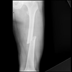
| | [[File:Femoral-shaft-fracture.jpeg|thumb|Femoral-shaft-fracture]] |
| | The [[femur]] is the largest and strongest [[bone]] in the body. Due to its strength it requires a significant force to break it. However, certain medical conditions that weaken the bone make it more vulnerable to fracture, so called [[Insufficiency Fracture|pathological fractures]]. For example: [[osteoporosis]]; [[Oncology|malignancy]]; [[Infectious Disease|infection]].<ref>Very well health Femur Fracture Available;https://www.verywellhealth.com/femur-fracture-2549281 (accessed 10.12.2022)</ref> |
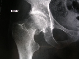
| | [[File:Neck of femur fracture (garden IV).jpeg|thumb|Neck of femur fracture]] |
|
| |
|
| A femoral fracture is a fracture of the [[femur]] (thigh bone). A femoral shaft fracture is defined as a fracture of the diaphysis occurring between 5 cm distal to the lesser trochanter and 5 cm proximal to the adductor tubercle occurs by chronic, repetitive activity that is common to runners and military. These injuries must be differentiated from insufficiency fractures, which, though similar in appearance and presentation, result from entirely different pathophysiology and occur in a different population.<ref>Michael S Wildstein, MD , Femoral Neck Stress and Insufficiency Fractures: http://emedicine.medscape.com/article/1246691-overview#a4 </ref> The femur is the largest and strongest bone in the body and has a good blood supply, so it requires a large or high impact force to break this bone.
| | == Location == |
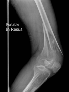
| | [[File:Distal-femoral-fracture.png|thumb|299x299px|Distal-femoral-fracture]] |
| | There are different types of femoral fractures according where they occur, namely: |
|
| |
|
| == Classification == | | # Femoral Head Fractures: often seen in the elderly osteoporotic population where the cortical bone is weak and so is the trabecular system. It occurs spontaneously or due to low-energy trauma. In the younger population, this fracture is rare and occurs due to high-energy trauma, usually associated with [[Hip Dislocation|hip dislocations]].<ref>Orthobullets Femoral Head Fractures Available:https://www.orthobullets.com/trauma/1036/femoral-head-fractures (accessed 10.12.2022)</ref> |
| | #[[Femoral Neck Hip Fracture|Femoral Neck Fractures]]: one of the most frequent fractures presenting to the emergency department and orthopedic trauma teams.<ref>[[Femoral Neck Hip Fracture|Hip Fracture]]</ref>See link for more. |
| | # [[Femoral Shaft Fractures]]: more common in men after a high-energy impact or in elderly women after a low-energy fall. <ref>Mercer's Textbook of Orthopaedics and Trauma Tenth edition edited by Suresh Sivananthan, Eugene Sherry, Patrick Warnke, Mark D Miller</ref> They can be described as follows: Type I - Spiral or transverse (most common) Type II – Comminuted Type III - Open <ref>WebMD. Broken bone: types of fractures, symptoms and prevention. 2014. (Available at http://www.webmd.boots.com/a-to-z-guides/bone-fractures-types-symptoms-prevention, accessed on 21 december 2014)fckLR</ref> |
| | # [[Distal femoral fracture]]: involve the femoral condyles and the metaphyseal region, often the resulting from high energy trauma eg motor vehicle accidents or a fall from a height. In the elderly, they may occur as an accident at home eg [[Falls in elderly|fall]].<ref>Radiopedia Distal femoral fracture. Available: https://radiopaedia.org/articles/distal-femoral-fracture (accessed 10.12.2022)</ref>See link for more. |
| | |
| | == Types == |
| There are 4 types of fracture: | | There are 4 types of fracture: |
| * Type 1: Stress fracture
| | # [[Femoral stress fracture]] |
| * Type 2: Severe impaction fractures
| | # Severe impaction fractures: the bone breaks into multiple fragments, which are driven into each other. It is a closed fracture that occurs when pressure is applied to both ends of the bone, causing it to split into two fragments that jam into each other. <ref>2.0 2.1 WebMD. Understanding Bone Fractures - the Basics. 2014. (Available at http://www.webmd.com/a-to-z-guides/understanding-fractures-basic-information, accessed 19 December 2014) fckLR</ref> |
| * Type 3: Partial fracture
| | # Partial fracture: incomplete break of a bone. This type of fracture refers to the way the bone breaks. In an incomplete fracture, the bone cracks but doesn’t break all the way through. In contrast, there is a complete fracture, where the bone snaps into two or more parts.<ref>Cleveland Clinic. Fractures. 2013. (Available at http://my.clevelandclinic.org/services/orthopaedics-rheumatology/diseases-conditions/hic-fractures, assessed 21 December 2014)fckLR</ref><ref>WebMD. Broken bone: types of fractures, symptoms and prevention. 2014. (Available at http://www.webmd.boots.com/a-to-z-guides/bone-fractures-types-symptoms-prevention, accessed on 21 december 2014)fckLR</ref> |
| * Type 4: Completed displaced fracture
| | # Completed displaced fracture: breaks into two or more pieces and is no longer correctly aligned. Displacement of fractures is defined in terms of the abnormal position of the distal fracture fragment in relation to the proximal bone. <ref>2.0 2.1 WebMD. Understanding Bone Fractures - the Basics. 2014. (Available at http://www.webmd.com/a-to-z-guides/understanding-fractures-basic-information, accessed 19 December 2014) fckLR</ref> |
| | |
| === According to the Severity of the Fracture: ===
| |
| This classification is for the severity of the fracture.
| |
| | |
| ==== Type 1: Stress Fracture ====
| |
| <br>A stress fracture is a small crack in the bone. Stress fractures often develop from overuse, such as from high-impact sports. Most stress fractures occur in the weight-bearing bones. A stress fracture is an overuse injury. When muscles are overtired, they are no longer able to lessen the shock of repeated impacts. When this happens, the muscles transfer the stress to the bones. This can create small cracks or fractures. <ref>AAOS. Stress Fractures of the Foot and Ankle. 2009. (Available at http://orthoinfo.aaos.org/topic.cfm?topic=a00379, accessed 19 December 2014) fckLR</ref>
| |
| | |
| [[File:Figuur_2.jpg|right|250x250px]]
| |
| | |
| ==== Type 2: Severe Impaction Fractures ====
| |
| <br>An impacted fracture is a fracture in which the bone breaks into multiple fragments, which are driven into each other. It is a closed fracture that occurs when pressure is applied to both ends of the bone, causing it to split into two fragments that jam into each other. <ref>2.0 2.1 WebMD. Understanding Bone Fractures - the Basics. 2014. (Available at http://www.webmd.com/a-to-z-guides/understanding-fractures-basic-information, accessed 19 December 2014) fckLR</ref> <br>
| |
| | |
| ==== Type 3: Partial Fracture ====
| |
| [[File:Figuur_3.jpg|right|250x250px]]
| |
| <br>A partial fracture is an incomplete break of a bone. This type of fracture refers to the way the bone breaks. In an incomplete fracture the bone cracks but doesn’t break all the way through. In contrast there is the complete fracture, where the bone snaps into two or more parts.<ref>Cleveland Clinic. Fractures. 2013. (Available at http://my.clevelandclinic.org/services/orthopaedics-rheumatology/diseases-conditions/hic-fractures, assessed 21 December 2014)fckLR</ref><ref>WebMD. Broken bone: types of fractures, symptoms and prevention. 2014. (Available at http://www.webmd.boots.com/a-to-z-guides/bone-fractures-types-symptoms-prevention, accessed on 21 december 2014)fckLR</ref><br>
| |
| | |
| ==== Type 4: Complete Displaced Fracture ====
| |
| [[File:Figuur_4.jpg|right|200x200px]]
| |
| <br>A bone has a displaced fracture when it breaks in two or more pieces and is no longer correctly aligned. Displacement of fractures is defined in terms of the abnormal position of the distal fracture fragment in relation to the proximal bone. <ref>2.0 2.1 WebMD. Understanding Bone Fractures - the Basics. 2014. (Available at http://www.webmd.com/a-to-z-guides/understanding-fractures-basic-information, accessed 19 December 2014) fckLR</ref><br>
| |
| === According to Location ===
| |
| Femoral fractures can be located at three different places:<br>
| |
| | |
| Femoral head fracture: Femoral head stress fractures are a common cause of hip pain in select populations. Chronic, repetitive activity that is common to runners and military recruits, predisposes these populations to femoral neck stress fractures. <ref>Michael S Wildstein, MD , Femoral Neck Stress and Insufficiency Fractures: http://emedicine.medscape.com/article/1246691-overview#a4 </ref>
| |
| | |
| <br> [[Image:Figuur 5.jpg|400x200px]]
| |
| | |
| <br>Source: Journal of the American Academy of Orthopaedic Surgeons<br>
| |
| | |
| ==== Head Fractures: The Pipkin Classification. ====
| |
| [[File:Figuur_6.jpg|right]]
| |
| <br>A: Type I, femoral head fracture inferior to the fovea centralis<br>B: Type II, fracture extended superior to the fovea centralis<br>C: Type III, any femoral head fracture with an associated femoral neck fracture<br>D: Type IV, any femoral head fracture with an associated acetabular fracture<br>
| |
| | |
| === Mechanism Of Injury ===
| |
| It is more often seen in the elderly population especially those who have senile osteoporosis wherein the cortical bone is weak and so is the trabecular system. It occurs spontaneously or due to low-energy trauma.
| |
| | |
| In the younger population, this fracture is rare and occurs due to high-energy trauma.
| |
| | |
| ==== Femoral Shaft Fracture ====
| |
| [[File:Enkel_titel_aangepast.jpg|right|350x350px]]
| |
| A femoral shaft fracture is defined as a fracture of the diaphysis occurring between 5 cm distal to the lesser trochanter and 5 cm proximal to the adductor tubercle. Femoral shaft fractures occur most frequently in young men after high-energy trauma and elderly women after a low-energy fall <ref>Mercer's Textbook of Orthopaedics and Trauma Tenth edition edited by Suresh Sivananthan, Eugene Sherry, Patrick Warnke, Mark D Miller</ref> Keany (2013) describes 3 types of femoral shaft fractures as follows: Type I - Spiral or transverse (most common) Type II – Comminuted Type III - Open <ref>WebMD. Broken bone: types of fractures, symptoms and prevention. 2014. (Available at http://www.webmd.boots.com/a-to-z-guides/bone-fractures-types-symptoms-prevention, accessed on 21 december 2014)fckLR</ref>
| |
| | |
| ==== Femoral Condyle Fracture ====
| |
| [[File:Figuur_7.jpg|right|300x300px]]
| |
| It occurs sometimes after a medial hamstring tendon ACL reconstruction with extra-articular tenodesis. The fracture occurs between the site of fixation of the extra-articular augmentation and the intraosseous femoral tunnel used in the intra-articular reconstruction <ref>Andrew R. J. Manktelow, Fares S. Haddad, FRCS (Orth), and Nicholas J. Goddard, FRCS . THE AMERICAN JOURNAL OF SPORTS MEDICINE, Vol. 26, No. 4.</ref><br>
| |
| | |
| == Clinically Relevant Anatomy ==
| |
| | |
| ==== Osteology ====
| |
| | |
| <br>The femur thigh bone, is the most proximal (closest to the hip joint) bone of the leg. The femur consists of a head, greater and lesser trochanter, shaft, and lateral and medial condyles with the patellar surface in between. The head of the femur articulates with the acetabulum in the pelvic bone forming the hip joint while the distal part of the femur articulates with the tibia and kneecap forming the knee joint. By most measures the femur is the strongest bone in the body. The femur is also the longest bone in the body. <ref>Keller K. et al. Muscle atrophy caused by limited mobilisation. 2013. </ref>
| |
| | |
| ==== Musculature ====
| |
| | |
| <br>The femur is the only bone in the thigh, it serves as an attachment point for all the muscles that exert their force over the hip and knee joints. Some bi-articular muscles, which cross two joints, like the [[gastrocnemius]] and [[plantaris]] muscles, also originate from the femur. <ref>Aukerman DF. Femur injuries and fractures. http://emedicine.medscape.com/article/90779-overview (accessed 30 October 2008) </ref>
| |
| | |
| *Quadriceps: [[Rectus Femoris|rectus femoris muscle]], [[Vastus Lateralis|vastus lateralis muscle]], [[Vastus Medialis|vastus medialis muscle]], [[Vastus Intermedius|vastus intermedius muscle]]
| |
| *[[Iliacus|Iliacus muscle]] (insertion on the lesser trochanter)
| |
| *[[Psoas Major|Psoas major muscle]] (Insertion on the lesser trochanter)
| |
| *Adductors: Adductor longus muscle, [[Adductor Brevis|adductor brevis muscle]], [[Adductor Magnus|adductor magnus muscle]], pectineus muscle, [[Gracilis|gracilis muscle]]
| |
| *[[Popliteus Muscle|Popliteus muscle]] (insertion under the lateral epicondyle)
| |
| *[[Gastrocnemius|Gastrocnemius muscle]] (Behind the adductor tubercle, over the lateral epicondyle and the popliteal facies)
| |
| *[[Plantaris|Plantaris muscle]] (over the lateral condyle)
| |
| *Abductors: [[Tensor Fascia Lata|tensor fasciae latae muscle]]
| |
| *Hamstrings: [[Biceps Femoris|biceps femoris muscle]], [[Semimembranosus|semimembranosus muscle]], [[Semitendinosus|semitendinosus muscle]]
| |
| *Gluteus muscles: insertion on the greater trochanter
| |
| *[[Piriformis|Piriformis muscle]]: insertion on the superior boundary of greater trochanter
| |
| *Gemellus superior muscle, gemellus inferior muscle, obturator internus muscle, obturator externus muscle, quadratus femoris muscle
| |
| | |
| <br>
| |
| | |
| #[[Image:Figuur 8.jpg|300x200px]]
| |
| | |
| Source picture: https://saveourbones.com/weekend-challenge-the-hip-bone-protector/
| |
| | |
| Following a femoral fracture, according to Keller et al., most of these muscles are much weaker than before, so physiotherapy is very important for muscle strengthening.
| |
| | |
| Several large muscles attach to the femur. Proximally, the gluteus medius and minimus attach to the greater trochanter, resulting in abduction of the femur with fracture. The iliopsoas attaches to the lesser trochanter, resulting in internal rotation and external rotation with fractures. The linea aspera (rough line on the posterior shaft of the femur) reinforces the strength and is an attachment for the gluteus maximus, adductor magnus, adductor brevis, vastus lateralis, vastus medialis, vastus intermedius, and short head of the biceps. Distally, the large adductor muscle mass attaches medially, resulting in an apex lateral deformity with fractures. The medial and lateral heads of the gastrocnemius attach over the posterior femoral condyles, resulting in Flexion deformity in distal-third fractures. <ref name="Keany">Keany, J. et al. Femur fracture. 2013. (Available at http://emedicine.medscape.com/article/824856-overview, accessed 6 November 2014)</ref> Also the dysfunction or atrophy of the muscles such as abductors, iliopsoas, adductors, gastrocnemius and tensor fascia lata can be an initiating point leading to femoral shaft fracture.
| |
| | |
| == Epidemiology/Etiology ==
| |
| | |
| Fractures of the femoral shaft are caused by a high-energy injury, such as a road traffic accident unless a pathological fracture in a patient with osteoporosis or metastatic disease. They are often associated injuries to the hip, pelvis, knee and other parts of the body. <br>Fractures vary in degree and complexity, depending on the degree of force involved. They may be: transverse (horizontally across the shaft), oblique, spiral (due to a twisting force), comminuted (when there are three or more resulting bone pieces), open or closed. <ref name="Keany" /><ref>Consensus Development Conference Prophylaxis and treatment of osteoporosis. American Journal of Medicine. 1991;90:107–110.</ref><br>The incidence of femoral fractures is reported as 1-1.33 fractures per 10,000 population per year in the USA. In the United States and Europe, the incidence of femoral fractures in children comprises 20 per 100,000 yearly. In individuals younger than 25 years and those older than 65 years, the rate of femoral fractures is 3 fractures per 10,000 population annually. <ref>Keller K. et al. Muscle atrophy caused by limited mobilisation. 2013.</ref>
| |
| | |
| Their incidence is in constant increase probably due to the demographic modifications and the continuous increment of the average life of the population and therefore the presence of a higher number of elderly patients. The reduction of BMD related to age is the main factor which exposes elderly people to a greater risk of hip fracture. Hip fractures are strongly associated with BMD in the proximal femur, but there are also many clinical predictors of hip fracture risk that are independent of bone density. Hip fracture incidence was 17 times greater among 15% of the women who had five or more of the risk factors, exclusive of bone density, compared with 47% of the women who had two risk factors or less. However, the women with five or more risk factors had an even greater risk of hip fracture if their bone density Z score was in the lowest tertile.]<ref>Consensus Development Conference Prophylaxis and treatment of osteoporosis. American Journal of Medicine. 1991;90:107–110. </ref><ref>Lars Kolmert, KristerWulf. Epidemiology and Treatment of Distal Femoral Fractures in Adults. </ref>
| |
| | |
| Several factors are related to increased risk of femoral fracture. Older persons (over 70) have a higher incidence of femoral fracture. A fracture of the proximal femoral is common in the elderly, with special emphasis on intracapsular (of the femoral neck) and extracapsular (trantrochanteric and subtrochanteric). Persons with Osteoporosis are also more likely to break their femur.<ref>7.0 7.1 Aukerman DF. Femur injuries and fractures. http://emedicine.medscape.com/article/90779-overview (accessed 30 October 2008)</ref>
| |
| | |
| The risk of these fractures increases exponentially with the increase of age and is higher in women (male-female ratio). Because women have more bone loss and fall than men, their incidence of hip fractures is about twice that seen in men at any age in the USA and Europe. Furthermore, women live longer than men so that more than three-quarters of all hip fractures occur in women. <ref>Cummings SR. Prevention of hip fractures in older women: a population-based perspective. Osteoporos Int. 1998;8(1):S8–12. </ref><ref>Kanis JA, Oden A, Johnell O, Johansson H, et al. The use of clinical risk factors enhances the performance of BMD in the prediction of hip and osteoporotic fractures in men and women. Osteoporos Int.2007;18(8):1033–46. </ref>
| |
| | |
| Morbidity and mortality rates have been reduced in femoral shaft fractures, mainly as the result of changes in methods of fracture immobilization. Current therapies allow for early mobilization, thus reducing the risk of complications associated with prolonged bed rest.
| |
| == Characteristics/Clinical Presentation ==
| |
| | |
| A broken thigh bone is almost always very obvious. Signs of a fracture include severe pain, inability to move the leg or stand on it, marked limitation of hip movements, local swelling and bruised skin. Typical for femoral neck and trochanteric fractures is the external rotation and the shortened lower limb, but both signs are less pronounced in a femoral neck fracture. Trochanteric fractures tend to cause more pain. Pain at the trochanteric area speaks in favour of a trochanteric fracture, whereas pain in the groin is typical of a neck fracture. <ref>Internal fixation of femoral neck fractures. An atlas. Manninger, Bosch, Cserháti, Fekete, Kazár. Eds Springer, 2007. </ref> <br>In neck or trochanteric fractures an anterior angulation at the fracture site, a recurvatum, may be present.<ref>WebMD. Broken bone: types of fractures, symptoms and prevention. 2014. (Available at http://www.webmd.boots.com/a-to-z-guides/bone-fractures-types-symptoms-prevention, accessed on 21 december2014)fckLR </ref> The absence of external rotation is rather seen in an undisplaced fracture, a shortened leg suggests the fracture is displaced. Yet limb shortening is not seen in fractures displaced in valgus and the limb might even be slightly longer. There may also be a resultant loss of blood in the femur and a haematoma may be present in the surrounding soft tissue. In case of an open fracture: open fractures have added the potential for infection.<ref>AAOS. Stress Fractures of the Foot and Ankle. 2009. (Available at http://orthoinfo.aaos.org/topic.cfm?topic=a00379, accessed 19 December 2014) fckLR</ref>Also knee ligament injuries are common, such as lateral collateral ligament injuries or anterior cruciate ligament injury and must be assessed after fracture fixation.<ref>10.0 10.1 Koval, K., & Zuckerman, J. et al. Handbook of Fractures. 2002; second edition. </ref>
| |
| | |
| == Differential Diagnosis ==
| |
| | |
| Hip pain can be localized to one of three anatomic regions. Pain associated with the anterior hip is mostly an intra-articular pathology (ex. Osteoarthritis and hip labral tears). Posterior hip pain can mostly be associated with piriformis syndrome, sacroiliac joint dysfunction, lumbar radiculopathy and ischio-femoral impingement and vascular claudication. The two last ones are less commonly. Greater trochanteric pain syndrome occurs when lateral hip pain is present. <ref>WILSON J.J. et al, Evaluation of the Patient with Hip Pain, American Academy of Family Physicians, January 2014, Volume 89, Number 1 </ref><ref>DeAngelis N.A. et al, Assessment and differential diagnosis of the painful hip, Clinical Orthopaedics& Related Research, January 2003, Volume 406, p. 11-18 </ref>
| |
| | |
| <br>Age can also distinguish the differential diagnosis of hip pain. Older patients are more associated with degenerative osteoarthritis and fractures. Congenital malformations of the femoro-acetabular joint, avulsion fractures, apophyseal or epiphyseal injuries are mostly found in prepubescent or adolescent patient. For adults or skeletally mature patients, hip pain is often related to musculotendinous strain, ligamentous sprain, contusion or bursitis. <ref>WILSON J.J. et al, Evaluation of the Patient with Hip Pain, American Academy of Family Physicians, January 2014, Volume 89, Number 1 </ref>
| |
| | |
| <br>When patients are asked about their hip pain several questions should be asked like antecedent trauma or inciting activity, factors that decrease/increase pain, mechanism of the injury and time of onset. <ref>WILSON J.J. et al, Evaluation of the Patient with Hip Pain, American Academy of Family Physicians, January 2014, Volume 89, Number 1 </ref><br>In the following table causes of hip pain are presented: <ref>Libor L.M. et al, Differential Diagnosis of Pain Around the Hip Joint, Arthroscopy: The Journal of Arthroscopic & Related Surgery, December 2008, Volume 24, Issue 12, Pages 1407–1421</ref>
| |
| | |
| Intra-articular:
| |
| | |
| *Labral tears
| |
| *Loose bodies
| |
| *Femoroacetabular impingement
| |
| *Capsular laxity
| |
| *Lig. Teres rupture
| |
| *Chondral damage<br>
| |
| Extra-articulair:
| |
| | |
| *Iliopsoas tendonitis
| |
| *Iliotibial band
| |
| *Gluteus medius/minimus
| |
| *Greater trochanteric bursitis
| |
| *Stress fracture
| |
| *Piriformis syndrome
| |
| *Sacroiliac joint pathology
| |
| | |
| Hip mimickers:
| |
| | |
| *Athletic pubalgia
| |
| *Sports hernia
| |
| *Osteitis pubis<br>
| |
| Livingston et al. also described the differential diagnosis for lateral hip pain <ref>Livingston J.I., Differential Diagnostic Process and Clincal Decision Making in a Young Adult Female With Lateral Hip Pain: A Case Report, The International Journal of Sports Physical Therapy, October 2015, Volume 10, Number 5 </ref>
| |
| | |
| *Acetabular labral tear
| |
| *Stress fracture, dislocation, fracture, contusion
| |
| *Osteonecrosis, avascular necrosis
| |
| *Muscle strain/tear, ligament sprain
| |
| *Low back pain, sacroiliac joint dysfunction
| |
| *Snapping hip syndrome
| |
| *Femoral acetabular impingement
| |
| *Bursitis Nerve entrapment syndrome
| |
| *Inflammatory disorders such as seronegative arthropathy, rheumatoid arthritis Infection
| |
| *Childhood disorders (Legg-Calve-Perthes Disease)
| |
| *Metabolic disease Tumor Primary or secondary osteoarthritis
| |
| *Psychosocial factors
| |
| == Diagnostic Procedures ==
| |
| | |
| On plain radiographs, anteroposterior (AP) and lateral views demonstrate most hip fractures. Check the neck-shaft angle, which is determined by measuring the angle created by lines drawn through the centers of the femoral shaft and femoral neck. This should be approximately 120-130°. If radiographic findings are equivocal but the history and physical examination are concerning for fracture, CT scan should be considered. One drawback to this modality, however, is that findings on scintigraphy are often negative during the first 24 hours after stress fracture. The positive predictive value of radionuclide imaging in diagnosing femoral neck stress pathology approaches 68%.<ref>Moira Davenport: Hip Fracture in Emergency Medicine, http://emedicine.medscape.com/article/825363-overview</ref><br>For patients in whom femoral neck fracture is strongly suspected but standard x-ray findings are negative, an AP view with internal rotation provides a better view of the femoral neck.
| |
| | |
| <br>If standard radiograph findings are negative and hip fracture still is strongly suspected, MRI and bone scan have high sensitivity in identifying occult injuries; MRI is 100% sensitive in patients with equivocal radiographic findings. For patients in whom a fracture is strongly suspected and radiographs are negative, consider admission if MRI or bone scan is not readily available. <ref>Michael S Wildstein, MD , Femoral Neck Stress and Insufficiency Fractures: http://emedicine.medscape.com/article/1246691-overview#a4 </ref>
| |
| | |
| == Outcome Measures ==
| |
| *[[Dynamic Gait Index|Dynamic Gait Index]]: The Dynamic Gait Index was developed as a clinical tool to assess gait, balance and fall risk. Because the DGI evaluates not only usual steady-state walking but also walking during more challenging tasks, it may be an especially sensitive test. <ref>Herman T, Inbar-Borovsky N, Brozgol M, Giladi N, Hausdorff JM. The Dynamic Gait Index in Healthy Older Adults: The Role of Stair Climbing, Fear of Falling and Gender. Gait& posture. 2009;29(2):237-241</ref>
| |
| *[[International Hip Outcome Tool (iHOT)]]: The International Hip Outcome Tool-33 (iHOT-33) is a self-administered outcome based on a Visual Analogue Scale response format designed for a young and active population with hip pathology. <ref>Ruiz-Ibán MA, Seijas R, Sallent A, et al. The international Hip Outcome Tool-33 (iHOT-33): multicenter validation and translation to Spanish. Health and Quality of Life Outcomes. 2015;13:62 </ref>
| |
| | |
| *[[Lower Extremity Functional Scale (LEFS)|Lower Extremity Functional Scale]] The Lower Extremity Functional Scale is a questionnaire containing 20 questions about a person's ability to perform everyday tasks. The LEFS can be used by clinicians as a measure of patients' initial function, ongoing progress and outcome, as well as to set functional goals.<<ref>Binkley JM, Stratford PW, Lott SA, Riddle DL. The Lower Extremity Functional Scale (LEFS): scale development, measurement properties, and clinical application. North American Orthopaedic Rehabilitation Research Network. Phys Ther. 1999 Apr;79(4):371-83.</ref>
| |
| | |
| * [[Timed Up and Go Test (TUG)]] A simple, quick and widely used clinical performance-based measure of lower extremity function, mobility and fall risk. We speculated that its properties may be different from other performance-based tests and assessed whether cognitive function may contribute to the differences among theses tests in a cohort of healthy older adults. <ref>Herman T, Giladi N, Hausdorff JM. Properties of the “Timed Up and Go” Test: More than Meets the Eye. Gerontology. 2011;57(3):203-210. </ref>
| |
| *A study suggests that the four-factor Mini-BESTest model can evaluate multiple dynamic balance aspects in older adults with femoral or vertebral fractures and may help therapists in making clinical decisions, considering factors that indicated a decline in function<ref>Miyata K, Hasegawa S, Iwamoto H, Otani T, Kaizu Y, Shinohara T, Usuda S. Comparison of the structural validity of three Balance Evaluation Systems Test in older adults with femoral or vertebral fracture. Journal of Rehabilitation Medicine. 2020 Jun 16.</ref>.
| |
| *[[Harris Hip Score]] is a validated tool and is usually used before and after hip surgery. It includes four domains like pain, functional status , deformity and range of motion.<ref name=":0">Gupta L, Lal M, Aggarwal V, Rathor LP. Assessing functional outcome using modified Harris hip score in patients undergoing total hip replacement. Int. J. Orthop. Sci. 2018 Apr 1;4:1015-7.</ref>
| |
| *Modified Harris Hip Score as the name suggests is the modified version of the Harris Hip Score. It includes the domains of pain, gait( limp, support and distance walked) and functional activities(Stairs, socks/shoes, public transportation, sitting). It is usually used after total hip replacement to assess their functional outcome.<ref>Kumar P, Sen R, Aggarwal S, Agarwal S, Rajnish RK. Reliability of Modified Harris Hip Score as a tool for outcome evaluation of Total Hip Replacements in Indian population. Journal of clinical orthopaedics and trauma. 2019 Jan 1;10(1):128-30.</ref>This scale is used preoperatively too. The only difference in this scale and the Harris Hip Score is that in the modified one the clinical evaluation part was removed.<ref name=":0" />
| |
| | |
| == Examination ==
| |
| | |
| It is important that your doctor know the specifics of how you hurt your leg. For example, if you were in a car accident, it would help your doctor to know how fast you were going, whether you were the driver or a passenger, whether you were wearing your seat belt, and if the airbags went off. This information will help your doctor determine how you were hurt and whether you may be hurt somewhere else. <ref>Brett Crist, MD; Stuart J. Fischer, MD; Stephen Kottmeier, MD.Femur Shaft Fractures (Broken Thighbone) (</ref><br>It is also important for your doctor to know whether you have other health conditions like high blood pressure, diabetes, asthma, or allergies. Your doctor will also ask you about any medications you take.
| |
| | |
| <br>After discussing your injury and medical history, the doctor carries out a careful examination. He or she will assess your overall condition and then focus on your leg. He will look for:
| |
| | |
| *An obvious deformity of the thigh/leg (an unusual angle, twisting, or shor,tening of the leg)
| |
| *Breaks in the skin
| |
| *Bruises
| |
| *Bony pieces that may be pushing on the skin<br>
| |
| After the visual inspection, your doctor will then feel along your thigh, leg, and foot looking for abnormalities and checking the tightness of the skin and muscles around your thigh. He or she will also feel for pulses. If you are awake, your doctor will test for sensation and movement in your leg and foot.
| |
| | |
| <br>The [[Ottawa Knee Rules]] are a clinical tool that can be used to determine the need for radiography following knee injury based on the patient's presentation. Other tests that will provide your doctor with more information about your injury include: <ref>Evans P.J., McGrory B.J. Fractures of the proximal femur. Hosp Physician. 2002:30–38. </ref>
| |
| | |
| *X-rays. The most common way to evaluate a fracture is with x-rays, which provide clear images of bone. [[X-Rays|X-rays]] can show whether a bone is intact or broken. They can also show the type of fracture and where it is located within the femur.
| |
| *Computed tomography (CT) scan. If your doctor still needs more information after reviewing your x-rays, he or she may order a [[CT Scans|CT scan]]. A CT scan shows a cross-sectional image of your limb. It can provide your doctor with valuable information about the severity of the fracture. For example, sometimes the fracture lines can be very thin and hard to see on an x-ray. A CT scan can help your doctor see the lines more clearly.
| |
| == Medical Management ==
| |
| | |
| Intracapsular and extracapsular fractures must be viewed as separate and distinct entities. The treatment of intracapsular fractures, which includes femoral head, subcapital or transcervical femoral neck fractures, must account for compromised blood flow and thus be geared toward its prostatic replacement, maintenance or restoration. <ref>Koval K.J. et al, Handbook of Fractures, Lippincott Williams & Wilkins, second edition, 2002, p. 180-181; 186-187; ev. </ref><ref>Grigoryan K.V .et al, Orthogeriatric care models and outcomes in hip fracture patients: a systematic review and meta-analysis. J Orthop Trauma, 2014;28(3): e49–e55. </ref><ref>Koval K.J. et al, Grigoryan K.V. et al,</ref> For extracapsular fractures, which include basicervical and intertrochanteric fractures, treatment focuses on reduction of the displacement and stabilization with implants for early mobilization and weight bearing. <ref>Handoll H.H.G. et al, Osteotomy, compression and other modifications of surgical techniques for internal fixation of extracapsular hip fractures (Review), Cochrane Library, 2009 </ref>
| |
| === Intracapsular Fractures ===
| |
| | |
| ==== Complete femoral head fractures ====
| |
| <br>Complete femoral head fractures are mostly located on the posterior side of the hip. Initial treatment consists of reduction, unregarded of the presence or type of femoral fracture. The risk of complication, like avascular necrosis, will decrease within the first few hours of injury when reduction is applied. <ref>Sheehan S.E. et al, Proximal Femoral Fractures: What the Orthopedic Surgeon Wants to Know, RadioGraphics, 2015, Volume 35-Number 5 </ref><br>In the majority of the cases, including dislocations of the femoral head and acetabulum, early closed reduction is favored. Yet, it is contraindicated when a femoral neck fracture is present.<ref>Loizou C.L. et al, Classification of subtrochanteric femoral fractures. Injury, 2010;41(7):739–745.</ref>
| |
| | |
| ==== Femoral Head Impaction Fractures ====
| |
| <br>The treatment of femoral head impaction fractures is controversial and challenging in younger patients. It is suggested, for elderly patients, to use a treatment consisting of reconstruction with total hip arthroplasty whereby damaged bone can be replaced. <ref>Sheehan S.E. et al, Proximal Femoral Fractures: What the Orthopedic Surgeon Wants to Know, RadioGraphics, 2015, Volume 35-Number 5 </ref>
| |
| '''<br>'''
| |
| | |
| ==== Femoral Neck Fractures ====
| |
| <br>Internal fixation with specific fixation approach depending on the pattern of the fracture is most often used to treat nondisplaced or impacted femoral neck fractures. Generally, they have good results for young and elderly patients. Early fixation is important to prevent the development of displacement of fractures. Which will happen in 10-30% if not treated. <ref>Sheehan S.E. et al, Proximal Femoral Fractures: What the Orthopedic Surgeon Wants to Know, RadioGraphics, 2015, Volume 35-Number 5 (</ref> A national pragmatic RCT: the '''SENSE''' trial will compare arthroplasties with internal fixation for undisplaced fracture neck femur in patients aged over 65<ref>Viberg B, Kold S, Brink O, Larsen MS, Hare KB, Palm H. [https://www.ncbi.nlm.nih.gov/pmc/articles/PMC7552868/ Is arthroplaSty bEtter than interNal fixation for undiSplaced femoral nEck fracture? A national pragmatic RCT: the SENSE trial]. BMJ open. 2020 Oct 1;10(10):e038442.</ref>.<br>For valgus/varus impacted injuries and Garden 2 fractures, the treatment consists of internal fixation with three cannulated lag screws. This also applies for Pauwels 1 and 2 fractures, but can alternately be fixed with sliding hip screw. Pauwels 3 fractures have a higher risk of instability and are therefore more problematic.<ref>Koval K.J. et al, Handbook of Fractures, Lippincott Williams & Wilkins, second edition, 2002, p. 180-181; 186-187; ev. </ref><ref>Unnanuntana A. et al, Atypical femoral fractures: what do we know about them?, AAOS Exhibit Selection. J Bone Joint Surg Am 2013, 95(2):e8 1–13. 71. </ref>
| |
| | |
| === Extracapsular Fractures ===
| |
| | |
| ==== ''' '''Intertrochanteric Fractures ====
| |
| <br>The treatment for intertrochanteric fractures consist of fixation using a laterale plate and screw fixation or intramedullary nail fixation. There is though no clear consensus regarding which implant is optimal for treating simple fracture patterns. <ref>Sheehan S.E. et al, Proximal Femoral Fractures: What the Orthopedic Surgeon Wants to Know, RadioGraphics, 2015, Volume 35-Number 5 </ref>
| |
| | |
| ==== ''' '''Subtrochanteric Fractures ====
| |
| <br>Displacement of the subtrochanteric includes flexion from iliopsoas muscle, abduction for gluteus medius and minimus muscles and external rotation from piriformis. An intramedullary nail is most used to achieve stability. Also often required is an open approach with direct reduction. <ref>Sheehan S.E. et al, Proximal Femoral Fractures: What the Orthopedic Surgeon Wants to Know, RadioGraphics, 2015, Volume 35-Number 5 </ref>
| |
| | |
| {{#ev:youtube|-5hE0Ambr5A|300}}
| |
| | |
| Video Animation of Intramedullary Nail Fixation (VIDEO)
| |
| | |
| Surgical reduction and fixation are indicated for the following types of proximal femur fractures:
| |
| | |
| *Intracapsular femoral neck fracture
| |
| *Dislocated femoral head
| |
| *Intertrochanteric fracture
| |
| *Subtrochanteric fracture <ref>Kisner C, Colby LA. Therapeutic exercises: foundations and techniques. Philadelphia: F.A. Davis, 2012.</ref>
| |
| '''Splints'''
| |
| | |
| <br>Alternatively, patients who are not medically stable or non ambulatory may be treated with traction. Applying a traction splint results in the reduction of hemorrhage. This is also one of the primary indications for applying these splints. The most common types of splints are the Thomas and Hare splint. Of which the Hare splint is more preferable because of the small application time. Femoral fractures need to be splinted during the initial phase. Those splints are needed to minimize pain and additional tissue damage.<br>These splints also have complications resulting from the use of traction splints including iatrogenic peroneal nerve injury, pressure sore development, ligamentous injury and pain. Even though these complications research had advised to apply significant forces because they are necessary to obtain fracture reduction. <ref>Daugherty, M. et al. Significant Rate of Misuse of the Hare Traction Splint for Children with Femoral Shaft Fractures. Journal of Emergency Nursing. 2013.fckLR</ref>[Daugherty, M. et al]<br>Surgical reduction and fixation should be performed within 24-48 hours of the injury.
| |
| | |
| == Physical Therapy Management ==
| |
| | |
| Whilst in hospital, a therapist will teach the patient how to use a walking aid to allow them to mobilise, depending on their weight bearing status. The patient should be taught basic range of movement and strengthening exercises to maintain a degree of strength and reduce the risk of blood clots.
| |
| | |
| Surgical fixation and immobilization are followed by extensive physical therapy. Under extensive therapy can be understood that the patients who underwent a femur fracture should receive a treatment by a physiotherapist who will invest time in gait training. Gait training results in increased bone formation. Even if gait training is completed using 30-50% of body weight support, an increase in bone formation could be found. <ref name="Carvalho et al.">Carvalho, D.C.L et al. Effect of treadmill gait on bone markers and bone mineral density of quadriplegic subjectshttp. 2006.</ref> After a femoral fracture, most of the muscles are much weaker than before so physiotherapy is very important. <br>
| |
| | |
| The physiotherapist will begin with a range of motion exercises for the hip, knee and ankle because mobility is decreased following immobilization. Mobilization is a very important treatment in the recovery process. The patient can also begin strengthening exercises based on the surgeon's orders (typically six weeks post-op). Patients should also undergo balance and proprioceptive rehab and these abilities are quickly lost with inactivity. <br>
| |
| | |
| ==== Mobility exercises ====
| |
| | |
| Knee: flexion and extension, abduction and adduction<br>Hip: flexion and extension, abduction and adduction, rotation <br> Functional quadriceps exercises should be initiated as soon as possible after the surgery because the quadriceps help provides stability in the knee. Flexion exercises also need to start as soon as possible, provided the fracture is adequated supported (i.e. the selected fixation approach allows for weight bearing). Physiotherapy should be continued until an acceptable functional range has been achieved or until a static position has been reached. It is necessary to record the range of movements in the knee with accuracy; first, this should be done at weekly and then at monthly intervals. <br>
| |
| | |
| During the postoperative treatment of patients with a proximal femoral fracture, physical therapy should focus on increasing the muscle strength, to improve walking safety and efficiency. Allowing the elderly patient to become more independent.
| |
| | |
| Research indicates that strengthening the abductor and adductor muscles of the hip increase the mediolateral stability during walks. Resulting in an influence on the improvement of the patient’s dynamic balance.<ref name="Carneiro et al.">Carneiro, M. et al. Physical therapy in the postoperative of proximal femur fracture in elderly. Literature review. 2013. fckLR(Levels of evidence: 1A)</ref><br>
| |
| | |
| ==== Orthopaedic therapy: ====
| |
| | |
| <br>During the immobilisatin period of the therapists need to actively mobilise the foot, with or without weight. Another important aspect is the mobilisation through a hole in the plaster of the patellar bone. The use of isometric exercises are also important to train the muscles (quadriceps, hamstring & glutei) of the upper leg. After the immobilisation period it is necessary to fixate the leg manually or by using a brace. The fixation is needed for the re-education of hip and knee and to secure a progressively verticalisation of the leg and to make the patient independent while walking or during other activities. Also, stabilisation exercises with unilateral support are recommended, but also balneotherapy. After the consolidation therapists need to focus on progressively increased pressure, the revalidation of the gait cycle, more intense mobilisation, strength-training therapy to reverse the muscle atrophy that occurred during the immobilisation period and condition training to increase the loss of endurance during the immobilisation period.<br>
| |
| ==== Osteosynthesis: ====
| |
| | |
| <br>Before going to bed, it is advised to massage the pre-articular structures and mobilisation of the hip. All mobilisation techniques can be used except rotations! It is also advised to use passive mobilisation techniques for retrieving the mobility of the patellar bone. The osteosynthesis is positively influenced by using isometric exercises of the hamstrings, quadriceps and glutei and the use of active exercises with low resistance to train the muscles of the hip and knee. The use of massage techniques of the quadriceps is advised straight after the verticalisation without support. The synthesis of the bone is also positively influenced by unilateral stabilisation exercises. After the consolidation period, orthopaedic therapy is recommended.<ref name="Xhardez et al.">Xhardez Y et al. Vade-mecum kinésithérapie et de reeducation fonctionnelle. 2002. fckLR(Levels of evidence: 5)</ref><br>
| |
| | |
| As non-operative treatment there are 3 alternatives:<br>1. Skin traction: used in adults only for emergency fracture immobilisation in the field for patient comfort and to facilitate patient transport. Main disadvantage is the causing of slippage or skin necrosis when applying sufficient forces to the limb.<br>2. Skeletal traction: used for early fracture care before a definitive operative procedure can be performed. The goal is to restore the femoral length and to limit rotational and angular deformities. Skeletal traction may be applied through the distal femur or proximal tibia. Problems with skeletal traction include knee stiffness, limb shortening, prolonged hospitalisation, respiratory and skin ailments, and malunion. <br>3. Cast Brace: an external support device that permits progressive weight bearing by partially unloading the fracture through circumferential support of the soft tissues. Indications for cast bracing include open fracture, distal third fractures, and comminuted midshaft fractures of the femur. Proximal, simple transverse, or oblique fractures are less amendable to cast bracing due to high stress concentration and a propensity to angulate. The cast brace is best used after an initial period of skeletal traction. Consistently high rates of union, superior to 90%, usually by 13 to 14 weeks, have been reported in numerous studies. Problems with cast bracing include loss of reduction and subsequent malunion, shortening and angulation.<ref name="Keany" /><br>
| |
| | |
| ==== Physical Exercise ====
| |
| | |
| <br>Improving muscle strength is necessary to enhance postoperative walking capacity for rehabilitation and to diminish the risks of falls. Physical activity will help:
| |
| | |
| *Preventing other fractures
| |
| *Increasing gait speed & balance
| |
| *Increasing ADL Performance
| |
| *Regaining walking capacity as early as possible after immobilization to avoid respiratory complications<ref>Mariana Barquet Carneiro, Débora Pinheiro Lédio Alves, and Marcelo Tomanik Mercadante. Physical therapy in the postoperative of proximal femur fracture in elderly. Literature review. ActaOrtopédicaBrasileira 2013 May-June; 21(3): 175–178. </ref>
| |
| *Better brain function and more social contact
| |
| | |
| Aerobic fitness is useful to include in a physical therapy plan for an improved cardiorespiratory capacity will lead to a better walking capacity. <br>Physical exercises are not only crucial for rehabilitation after fracture but for ongoing reinforcing of the mineral bone density, especially in vulnerable populations like elder fragile patients, osteoporotic post-menopausal women or people suffering from osteoporosis or osteopenia. Long-term odd-impact exercise-loading, is associated, similar to high-impact exercise-loading, with a 20% thicker cortex around the femoral neck <ref>Nikander R, Kannus P, Dastidar P, Hannula M, Harrison L, Cervinka T, Narra NG, Aktour R, Arola T, Eskola H, Soimakallio S, Heinonen A, Hyttinen J, Sievänen H. Targeted exercises against hip fragility. Osteoporos Int. 2009 Aug;20(8):1321-8.</ref>. In aerobic fitness, these type of movements are most frequently used.
| |
| | |
| Moderate magnitude impacts from Odd-exercise loading is mechanically less demanding and makes the body work in all directions, retraining the biomechanical qualities and properties of the bone structure and impacting positively the bone mineral density. Fitness aerobics, dance fitness, dance, ball games, gymnastics involving rapid turns and movements are good examples of odd-impact exercises.<br>Futhermore, duration, frequency and intensity are important and should be customized to the different age-groups.
| |
| | |
| Strengthening exercises seem to be key for the functional improvement. <ref>Mariana Barquet Carneiro, Débora Pinheiro Lédio Alves, and Marcelo Tomanik Mercadante. Physical therapy in the postoperative of proximal femur fracture in elderly. Literature review. ActaOrtopédicaBrasileira 2013 May-June; 21(3): 175–178.</ref> These strength exercises may as well produce advantages in the psychosocial area which tends to be altered in elder patients that suffered a fracture. Weight-bearing exercises will reinforce the dynamic balance and functional performance <ref>SD Yang, SH Ning, LH Zhang, YZ Zhang, WY Ding. The effect of lower limb rehabilitation gymnastics on postoperative rehabilitation in elderly patients with femoral shaft fracture: A retrospective case-control study. Medicine, 2016 - journals.lww.com </ref>, especially exercises in standing position since they are more challenging for the postural control <ref>Mariana Barquet Carneiro, Débora Pinheiro Lédio Alves, and Marcelo Tomanik Mercadante. Physical therapy in the postoperative of proximal femur fracture in elderly. Literature review. ActaOrtopédicaBrasileira 2013 May-June; 21(3): 175–178. </ref>.<br>
| |
| | |
| ==== Home Rehabilitation Training ====
| |
| | |
| <br>Home Rehabilitation Training leads towards better rehabilitation and better performance in daily activities. Home physiotherapy training is suitable for all elder patients, including those suffering from cognitive or psychological impairment. The literature stresses the importance of home physiotherapy, in combination with day-to-day activities like going to the shop, for gaining confidence, balance, functionality and reducing as such the number of falls. Therefore it can also be seen as a way to prevent falls. <ref>Andrea Giusti, MD, Antonella Barone, MD, Mauro Oliveri, MD, Monica Pizzonia, MD, Monica Razzano, MD, Ernesto Palummeri, MD, Giulio Pioli, MD, PhD. An Analysis of the Feasibility of Home Rehabilitation Among Elderly People With Proximal Femoral Fractures. Archives of Physical Medicine and Rehabilitation. June 2006, Volume 87, Issue 6, Pages 826-831.</ref>Fall prevention programmes are important for the elder population that already suffers a femoral fracture. The literature indicates that elder people often fall again following a previous hip or femoral fracture and that this constitutes a major health problem <ref>M. Berggren, M. Stenvall, B. Olofsson, Y. Gustafson.Osteoporosis International . Evaluation of a fall-prevention program in older people after femoral neck fracture: a one-year follow-up. June2008, Volume 19, Issue 6, pp 801-809. </ref>. The study of Berggren et al. concludes that fall-prevention must be part of everyday life in fall-prone elderly. Prevention and treatment of fall-risk factors are key. These programs should include gait training with advice on assistive devices and medication, exercise programmes for balance training, treatments for cardiovascular problems, environmental modifications and hypotension.<ref>M. Stenvall, B. Olofsson, M. Lundström, U. Englund, B. Borssén, O. Svensson, L. Nyberg, and Y. Gustafson. A multidisciplinary, multifactorial intervention program reduces postoperative falls and injuries after femoral neck fracture. Osteoporosis International. Feb 2007; 18(2): 167–175</ref>.
| |
| | |
| ==== Electrical Stimulation ====
| |
| | |
| <br>The literature is still not conclusive on this topic and the results of one study may contradict or, on the contrary, reinforce the results of another study. Yet there is evidence supporting the beneficial effects of electrical stimulation, especially in combination with physical therapy exercises. In a randomized controlled trial Gremeaux et al.conclude that « Low-frequency electric muscle stimulation can thus be proposed as a simple, effective, and safe complementary therapy used in conjunction with standard rehabilitation in everyday clinical practice in these patients.
| |
| | |
| In a critical literature review of 2005 Bax et al. stated that only limited evidence suggests that neuromuscular electrical stimulation can be more effective than no exercice in individuals with impaired and unimpaired quadriceps, and volitional exercises appeared more effective in most situations <ref>Braid V, Barber M, Mitchell SL, Martin BJ, Granat M, Stott DJ. Randomised controlled trial of electrical stimulation of the quadriceps after proximal femoral fracture AgingClinExp Res. 2008 Feb;20(1):62-6. </ref>. Low-frequency ES may lead to a significant increase in muscle strength in the operated limb and is overall well tolerated by the elder population.
| |
| | |
| <br>The terms « Functional electrical Stimulation », « Cyclic Stimulation » or « Neuromuscular Electrical Stimulation » all refer to the same « Electrical Stimulation » but speaking of it, distinction should be made between low-frequency and high-frequency stimulation. High-frequency stimulation acts principally on fast-twitch fibers, increasing muscle strength and resistance to fatigue. It could be useful to fight muscle de-conditioning but it is mostly not well tolerated in older patients.
| |
| | |
| <br>Low-frequency Electrical Stimulation (LFES) increases the metabolic activity of slow-twitch muscle fibers and the proportion of slow-twitch fibers. This fiber-type is more dominantly present in the Quadriceps and therefore interesting to use this low frequency on the knee-extensor muscle. Other structural adaptations are the development of mitochondrial apparatus and increase in capillary density, resulting in increased resistance to fatigue.<ref>Vincent Gremeaux, MD, Julien Renault, MS, Laurent Pardon, MS, GaelleDeley, PhD, Romuald Lepers, PhD, Jean-Marie Casillas, MD. Low-Frequency Electric Muscle Stimulation Combined With Physical Therapy After Total Hip Arthroplasty for Hip Osteoarthritis in Elderly Patients: A Randomized Controlled Trial Archives of Physical Medicine and Rehabiliation. December 2008, Volume 89, Issue 12, Pages 2265-2273. </ref>. LFES has also been used in the treatment of different neurologic and orthopedic disorders.
| |
| | |
| <br>ES may attenuate muscle atrophy, helps regaining muscle strength faster after surgery and show a trend towards improved autonomy and walking speed <ref>Vincent Gremeaux, MD, Julien Renault, MS, Laurent Pardon, MS, GaelleDeley, PhD, Romuald Lepers, PhD, Jean-Marie Casillas, MD. Low-Frequency Electric Muscle Stimulation Combined With Physical Therapy After Total Hip Arthroplasty for Hip Osteoarthritis in Elderly Patients: A Randomized Controlled Trial Archives of Physical Medicine and Rehabiliation. December 2008, Volume 89, Issue 12, Pages 2265-2273.</ref>. The results of the study of Gremeaux et al. suggest that low-frequency electric muscle stimulation associated with conventional physiotherapy is superior to physiotherapy alone in increasing strength of the knee extensors, which is accompanied by a return to better muscular equilibrium between the operated and non-operated limb. It leads to a significant increase in muscle strength in the operated limb. In young subjects ES can produce powerful muscle contraction and give training effects as good as or better than voluntary isometric exercise<ref>Braid V, Barber M, Mitchell SL, Martin BJ, Granat M, Stott DJ. Randomised controlled trial of electrical stimulation of the quadriceps after proximal femoral fracture AgingClinExp Res. 2008 Feb;20(1):62-6. </ref>
| |
| == Resources ==
| |
| | |
| *http://ac.els-cdn.com/S1878764914706291/1-s2.0-S1878764914706291-main.pdf?_tid=65bdb594-5516-11e4-8d24-00000aab0f01&acdnat=1413451721_842b7d8bdef872019e2b5a7408aca409/<br>
| |
| *http://apps.webofknowledge.com.ezproxy.vub.ac.be:2048/full_record.do?product=WOS&search_mode=Refine&qid=4&SID=S2zn1kgocpk8Xkj2bxO&page=1&doc=1
| |
| *http://www.scielo.br/scielo.php?script=sci_arttext&pid=S0100-879X2006001000012&lng=en&nrm=iso&tlng=en
| |
| *http://orthoinfo.aaos.org/topic.cfm?topic=a00379<br>
| |
| *http://www.houstonspineandjoint.com/bone-fracture-types.html<br>
| |
| *http://my.clevelandclinic.org/services/orthopaedics-rheumatology/diseases-conditions/hic-fractures
| |
| *http://www.ncbi.nlm.nih.gov/pubmed/23519955<br>
| |
| | |
| == References == | | == References == |
|
| |
|


