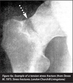
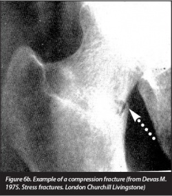
Femoral stress fracture: Difference between revisions
Evan Thomas (talk | contribs) mNo edit summary |
No edit summary |
||
| Line 7: | Line 7: | ||
'''Stress fractures''' are injuries that occur when repetitive and excessive stress on a [[Bone|bone]] is combined with limited rest. This leads to muscle weakness and a lower shock absorbing capacity of the leg<ref name="Stress">Stress Fractures, information from your family doctor. Americain Family Physician Jan. 2011. Level of evidence: 5</ref><ref name="Zadpoor">Zadpoor A, Nikooyan A. The relationship between lower-extremity stress fractures and the ground reaction force: a systematic review. Clinical Biomechanics 2011; 26: 23 -28. Level of evidence: 1B</ref><ref name="Niva">Niva M, Mattila V, Kiuru M, Pihlajamäki H. Bone stress Injuries are common in female military trainees. Clin Orthop Relat Res (2009) 467: 2962-2969. Level of evidence: 3A</ref><ref name="Schultz">Schultz, Houglum, Perrin. Third edition, examination of musculoskeletal injuries p.401. Human Kinetics. Level of evidence: 5</ref><ref name="Houglum">Houglum P. Second edition, therapeutic exercise for musculoskeletal injuries p.80-81, p. 811-812. Human Kinetics. Level of evidence: 5</ref>. At the begining there is only pain during activity, but in the next phase the pain also occurs after activity and during the night. There is a grading system of five grades based on MRI results, but this system is not yet validated. Grades I to III are the low grades <ref name="Niva" />, begining with endosteal edema followed by periosteal edema to muscle edema. When the injury reaches level IV, a fracture line can be observed on the imaging. A grade V refers to callus formation in the cortical bone <ref name="Niva" /><ref name="Fredericson">Fredericson M, Jang K, Bergman G, Gold G. Femoral diaphyseal stress fractures: results of a systematic bone scan and magnetic resonance evaluation in 25 runners. Physical Therapy in Sport 5 (2004): 188-193. Level of evidence: 2B</ref>. | '''Stress fractures''' are injuries that occur when repetitive and excessive stress on a [[Bone|bone]] is combined with limited rest. This leads to muscle weakness and a lower shock absorbing capacity of the leg<ref name="Stress">Stress Fractures, information from your family doctor. Americain Family Physician Jan. 2011. Level of evidence: 5</ref><ref name="Zadpoor">Zadpoor A, Nikooyan A. The relationship between lower-extremity stress fractures and the ground reaction force: a systematic review. Clinical Biomechanics 2011; 26: 23 -28. Level of evidence: 1B</ref><ref name="Niva">Niva M, Mattila V, Kiuru M, Pihlajamäki H. Bone stress Injuries are common in female military trainees. Clin Orthop Relat Res (2009) 467: 2962-2969. Level of evidence: 3A</ref><ref name="Schultz">Schultz, Houglum, Perrin. Third edition, examination of musculoskeletal injuries p.401. Human Kinetics. Level of evidence: 5</ref><ref name="Houglum">Houglum P. Second edition, therapeutic exercise for musculoskeletal injuries p.80-81, p. 811-812. Human Kinetics. Level of evidence: 5</ref>. At the begining there is only pain during activity, but in the next phase the pain also occurs after activity and during the night. There is a grading system of five grades based on MRI results, but this system is not yet validated. Grades I to III are the low grades <ref name="Niva" />, begining with endosteal edema followed by periosteal edema to muscle edema. When the injury reaches level IV, a fracture line can be observed on the imaging. A grade V refers to callus formation in the cortical bone <ref name="Niva" /><ref name="Fredericson">Fredericson M, Jang K, Bergman G, Gold G. Femoral diaphyseal stress fractures: results of a systematic bone scan and magnetic resonance evaluation in 25 runners. Physical Therapy in Sport 5 (2004): 188-193. Level of evidence: 2B</ref>. | ||
<br> | |||
[[Image:Femoral stress fracture tension.jpg|250px]] <ref name="Devas">Devas, Michael. Stress fractures. Churchill Livingstone, 1975.</ref>[[Image:Femoral stress fracture compression.jpg|250px]]<ref name="Devas" /> | |||
== Clinically Relevant Anatomy == | == Clinically Relevant Anatomy == | ||
| Line 49: | Line 53: | ||
*Bone marrow edema | *Bone marrow edema | ||
Therapists have to pay attention to misdiagnose a femoral stress fracture as [[ | Therapists have to pay attention to misdiagnose a femoral stress fracture as [[Muscle_Strain|muscle strain ]]of the quadriceps or illiopsoas tendinopathy because of the similar symptoms <ref name="Nguyen">Nguyen J, Peterson J, Biswal S, Beaulieu C, Fredericson M. Stress-related injuries around the lesser trochanter in long-distance runners. AJR 2008; 190: 1616-1620. Level of evidence: 3B</ref>.<br>To present femoral stress fractures on images. We can use 4 modalities: plain radiography, bone scintigrapy, MRI and ultrasonography with the highest sensitivity and specificity for MRI <ref name="Patel" />. These techniques can be used in different phases of diagnosis and treatment <ref name="Patel" /><ref name="Korvala" />.<br> | ||
== Examination<ref>Casterline M, Osowski S, Ulrich G. Femoral stress fracture. Journal of Athletic Training March 1996; 31: 53-56. Level of evidence: 4</ref> == | == Examination<ref>Casterline M, Osowski S, Ulrich G. Femoral stress fracture. Journal of Athletic Training March 1996; 31: 53-56. Level of evidence: 4</ref> == | ||
| Line 61: | Line 65: | ||
Initial treatment is based on the reduction of activities to a pain-free level. During this “relative rest”-period of 4 to 12 weeks, the patient can use pneumatic compression walking boots to reduce his pain level. Also physical therapy and cross-training which contains of flexibility, strength and cardiovascular training is permitted for example swimming and biking. After that restriction period the activities can be increased in a slow, graduated way <ref name="Snyder" />. Ivkovic et al. designed a new treatment algorithm for femoral shaft stress injuries. Four phases has to be fulfilled to start normal training and each phase is evaluated by a hop or fulcrum test. The first phase is called symptomatic, where the patient has to walk with crutches. The second phase is the asymptomatic one where patient are allowed to walk normally and to start swimming and exercises the upper extremity. During the third ‘basic’ phase the patient can perform exercises of lower and upper extremities. During the last ‘resuming phase’, the athlete is allowed to gradually start normal training <ref name="Ivkovic" />. No recurrence of injury after treatment and follow-up for 48-96 months <ref name="Ivkovic" />.<br>The treatment algorithm is free available in the article from Ivkovic et al.: "Stress fractures of the femoral shaft in athletes: a new treatment algorithm."<br><br>To prevent femoral stress fractures, people could modify their training schedules and wear shock-absorbing shoe inserts. Insoles lowers the incidence because the improves biomechanics, less fatigue and limit the impact on the ground. The size of these insoles can range in different types to support the forefoot and/or the toes <ref name="Zadpoor" /><ref name="Snyder" />. Also calcium and vitamin D supplementation could play a role in the prevention but their data are controversial <ref name="Snyder" />. Leg muscle stretching during warm-up has no significant effect on prevention for femoral stress fractures<ref name="Patel" />.<br> | Initial treatment is based on the reduction of activities to a pain-free level. During this “relative rest”-period of 4 to 12 weeks, the patient can use pneumatic compression walking boots to reduce his pain level. Also physical therapy and cross-training which contains of flexibility, strength and cardiovascular training is permitted for example swimming and biking. After that restriction period the activities can be increased in a slow, graduated way <ref name="Snyder" />. Ivkovic et al. designed a new treatment algorithm for femoral shaft stress injuries. Four phases has to be fulfilled to start normal training and each phase is evaluated by a hop or fulcrum test. The first phase is called symptomatic, where the patient has to walk with crutches. The second phase is the asymptomatic one where patient are allowed to walk normally and to start swimming and exercises the upper extremity. During the third ‘basic’ phase the patient can perform exercises of lower and upper extremities. During the last ‘resuming phase’, the athlete is allowed to gradually start normal training <ref name="Ivkovic" />. No recurrence of injury after treatment and follow-up for 48-96 months <ref name="Ivkovic" />.<br>The treatment algorithm is free available in the article from Ivkovic et al.: "Stress fractures of the femoral shaft in athletes: a new treatment algorithm."<br><br>To prevent femoral stress fractures, people could modify their training schedules and wear shock-absorbing shoe inserts. Insoles lowers the incidence because the improves biomechanics, less fatigue and limit the impact on the ground. The size of these insoles can range in different types to support the forefoot and/or the toes <ref name="Zadpoor" /><ref name="Snyder" />. Also calcium and vitamin D supplementation could play a role in the prevention but their data are controversial <ref name="Snyder" />. Leg muscle stretching during warm-up has no significant effect on prevention for femoral stress fractures<ref name="Patel" />.<br> | ||
== Resources == | == Resources == | ||
*Pubmed, Web of Knowledge, Pedro | *Pubmed, Web of Knowledge, Pedro | ||
*Third edition, Examination of musculoskeletal injuries | *Third edition, Examination of musculoskeletal injuries | ||
*Second edition, Therapeutic exercise for musculoskeletal injuries<br> | *Second edition, Therapeutic exercise for musculoskeletal injuries<br> | ||
== Recent Related Research (from [http://www.ncbi.nlm.nih.gov/pubmed/ Pubmed]) == | == Recent Related Research (from [http://www.ncbi.nlm.nih.gov/pubmed/ Pubmed]) == | ||
| Line 73: | Line 77: | ||
<references /><br> | <references /><br> | ||
[[Category:Condition]] [[Category:Bones]] [[Category:Hip]] [[Category:Knee]] [[Category:Sports_Injuries]][[Category:Musculoskeletal/Orthopaedics]] | [[Category:Condition]] [[Category:Bones]] [[Category:Hip]] [[Category:Knee]] [[Category:Sports_Injuries]] [[Category:Musculoskeletal/Orthopaedics]] | ||
Revision as of 20:18, 9 January 2017
Original Editors - Matthias Verstraelen as part of the Vrije Universiteit Brussel's Evidence-based Practice project
Top Contributors - Matthias Verstraelen, Lucinda hampton, Kim Jackson, Redisha Jakibanjar, Admin, Daniele Barilla, Adam Vallely Farrell, Wanda van Niekerk, Daphne Jackson, Laura Ritchie, Evan Thomas, Naomi O'Reilly, WikiSysop, Elise Audiens and Claire Knott
Definition/Description[edit | edit source]
Stress fractures are injuries that occur when repetitive and excessive stress on a bone is combined with limited rest. This leads to muscle weakness and a lower shock absorbing capacity of the leg[1][2][3][4][5]. At the begining there is only pain during activity, but in the next phase the pain also occurs after activity and during the night. There is a grading system of five grades based on MRI results, but this system is not yet validated. Grades I to III are the low grades [3], begining with endosteal edema followed by periosteal edema to muscle edema. When the injury reaches level IV, a fracture line can be observed on the imaging. A grade V refers to callus formation in the cortical bone [3][6].
Clinically Relevant Anatomy[edit | edit source]
Stress fractures of the femur can occur in the whole bone like the neck, shaft and the condyles. The highest incidence is seen at the femoral neck. When the patient doesn’t adapt his or her training, certain stress fractures could lead to complications, even to the point of complete femoral fractures of the head or shaft [8][9].
Epidemiology/Etiology[edit | edit source]
Stress fractures are presented in athletes with the main focus on running or in military trainees[3]. The femoral ones are at a level 6% to 7% of all the stress injuries. Femoral stress fractures are seen in overtrainers but also in undertrainers [10]. With the decreased physical level of the population, it is possible that the incidence of femoral stress fractures will increase in the future [6].
Characteristics/Clinical Presentation[edit | edit source]
The risk factors are as follows: [11][1][2][12]
- High-intensity training
- Recreational runners
- Track and field, basketball, soccer, dance
- Women
- Poor nutrition and lifestyle activities
- Lower 25-hydroxyvitamin D
- Female athlete triad
- (history of) smoking
- < 3 times exercising/week
- > 10 alcoholic drinks/week
- Genetic factors [13] (CTR C allele, VDR C-A haplotype, LRP5 A-G-G-C, VDR C-A haplotype)
- Change of surfaces (indoor track, frozen field)
- Biomechanical imbalance (leg length, foot arch, forefoot varus, stance of foot and ankle)
There is a trend to significance for energy expenditure (kcal/day) with lower limb stress fractures (p=0.06). This effect could be explained as a sequence that more active people have also a higher expenditure level [14].
No significant effect is found of the ground reaction force on the incidence of lower-limb stress fractures (p>0.05) [2].
The incidence of femoral stress fractures is reduced with 14% (p=0.013) when a semi rigid insole is used [15].
Alana et al. concluded that there’s no effect on lower limb stress fractures and calcium intake (p=0.55) or bone density [14].
There is a trend to significance for energy expenditure (kcal/day) with lower limb stress fractures (p=0.06). This effect could be explained as a sequence that more active people have also a higher expenditure level [14].
Diagnostic Procedures[edit | edit source]
First of all we describe the possible symptoms: [12][3][16][4]
- Local pain and edema
- Point tenderness on palpation
- Local swelling
- Antalgic gait
- Painful and limited passive and active ROM of hip and/or knee (flexion, internal rotation, extension)
- Pain increases during activity
- Groin pain
- Bone marrow edema
Therapists have to pay attention to misdiagnose a femoral stress fracture as muscle strain of the quadriceps or illiopsoas tendinopathy because of the similar symptoms [17].
To present femoral stress fractures on images. We can use 4 modalities: plain radiography, bone scintigrapy, MRI and ultrasonography with the highest sensitivity and specificity for MRI [11]. These techniques can be used in different phases of diagnosis and treatment [11][13].
Examination[18][edit | edit source]
The hop test and tuning fork test could be used as diagnostic test but there is a lack of recent evidence for their validity. Another test is the “fist” test, the therapist create a bilateral pressure on the anterior side of the femur starting at the distal part and moving to the proximal one. The most valid test for the diagnosis is the fulcrum-test, while the therapist pushes to the dorsum of the knee [16].
Physical Therapy Management[edit | edit source]
Initial treatment is based on the reduction of activities to a pain-free level. During this “relative rest”-period of 4 to 12 weeks, the patient can use pneumatic compression walking boots to reduce his pain level. Also physical therapy and cross-training which contains of flexibility, strength and cardiovascular training is permitted for example swimming and biking. After that restriction period the activities can be increased in a slow, graduated way [15]. Ivkovic et al. designed a new treatment algorithm for femoral shaft stress injuries. Four phases has to be fulfilled to start normal training and each phase is evaluated by a hop or fulcrum test. The first phase is called symptomatic, where the patient has to walk with crutches. The second phase is the asymptomatic one where patient are allowed to walk normally and to start swimming and exercises the upper extremity. During the third ‘basic’ phase the patient can perform exercises of lower and upper extremities. During the last ‘resuming phase’, the athlete is allowed to gradually start normal training [16]. No recurrence of injury after treatment and follow-up for 48-96 months [16].
The treatment algorithm is free available in the article from Ivkovic et al.: "Stress fractures of the femoral shaft in athletes: a new treatment algorithm."
To prevent femoral stress fractures, people could modify their training schedules and wear shock-absorbing shoe inserts. Insoles lowers the incidence because the improves biomechanics, less fatigue and limit the impact on the ground. The size of these insoles can range in different types to support the forefoot and/or the toes [2][15]. Also calcium and vitamin D supplementation could play a role in the prevention but their data are controversial [15]. Leg muscle stretching during warm-up has no significant effect on prevention for femoral stress fractures[11].
Resources[edit | edit source]
- Pubmed, Web of Knowledge, Pedro
- Third edition, Examination of musculoskeletal injuries
- Second edition, Therapeutic exercise for musculoskeletal injuries
Recent Related Research (from Pubmed)[edit | edit source]
References[edit | edit source]
- ↑ 1.0 1.1 Stress Fractures, information from your family doctor. Americain Family Physician Jan. 2011. Level of evidence: 5
- ↑ 2.0 2.1 2.2 2.3 Zadpoor A, Nikooyan A. The relationship between lower-extremity stress fractures and the ground reaction force: a systematic review. Clinical Biomechanics 2011; 26: 23 -28. Level of evidence: 1B
- ↑ 3.0 3.1 3.2 3.3 3.4 Niva M, Mattila V, Kiuru M, Pihlajamäki H. Bone stress Injuries are common in female military trainees. Clin Orthop Relat Res (2009) 467: 2962-2969. Level of evidence: 3A
- ↑ 4.0 4.1 Schultz, Houglum, Perrin. Third edition, examination of musculoskeletal injuries p.401. Human Kinetics. Level of evidence: 5
- ↑ Houglum P. Second edition, therapeutic exercise for musculoskeletal injuries p.80-81, p. 811-812. Human Kinetics. Level of evidence: 5
- ↑ 6.0 6.1 Fredericson M, Jang K, Bergman G, Gold G. Femoral diaphyseal stress fractures: results of a systematic bone scan and magnetic resonance evaluation in 25 runners. Physical Therapy in Sport 5 (2004): 188-193. Level of evidence: 2B
- ↑ 7.0 7.1 Devas, Michael. Stress fractures. Churchill Livingstone, 1975.
- ↑ Patel D, Roth M, Kapil N. Stress fractures: diagnosis, treatment and prevention. American Family Physician Jan. 2011; 83: 39-46. Level of evidence: 1A
- ↑ Zadpoor A, Nikooyan A. The relationship between lower-extremity stress fractures and the ground reaction force: a systematic review. Clinical Biomechanics 2011; 26: 23 -28. Level of evidence: 1B
- ↑ Kang L, Belcher D, Hulstyn M. Stress fractures of the femoral shaft in women’s college lacrosse: a report of seven cases and a review of the literature. Br J Sports Med 2005; 39: 902-906. Level of evidence: 2B
- ↑ 11.0 11.1 11.2 11.3 Patel D, Roth M, Kapil N. Stress fractures: diagnosis, treatment and prevention. American Family Physician Jan. 2011; 83: 39-46. Level of evidence: 1A
- ↑ 12.0 12.1 Anand A, Raviraj A, Kodikal G. Subchondral stress fractures of femoral head in healthy adult. Indian J Orthop. 2010 Oct-Dec; 44(4): 458-460. Level of evidence: 3B
- ↑ 13.0 13.1 Korvala J, Hartikka H, Pihlajamäki H, Solovieva S, Ruohola J-P, Sahi T, Barral S, Ott J, Ala-Kokko L, Männikkö M. Genetic predisposition for femoral neck stress fractures in military conscripts. BMC Genetics 2010, 11: 95. Level of evidence: 2B
- ↑ 14.0 14.1 14.2 Cline A, Jansen R, Melby C. Stress fractures in female army recruits: implications of bone density, calcium intake and exercise. Journal of the American College of Nutrition, Vol. 17; No. 2: 128-135 (1998). Level of evidence: 3A
- ↑ 15.0 15.1 15.2 15.3 Snyder R, De Angelis J, Koester M., Spindler K, Dunn W. Does shoe insole modification prevent stress fractures? A systematic review. HSSJ (2009) 5: 92-98. Level of evidence: 2B
- ↑ 16.0 16.1 16.2 16.3 Ivkovic A, Bojanic I, Pecina M. Stress fractures of the femoral shaft in athletes: a new treatment algorithm. Br J Sports Med 2006; 40: 518-520. Level of evidence: 2A
- ↑ Nguyen J, Peterson J, Biswal S, Beaulieu C, Fredericson M. Stress-related injuries around the lesser trochanter in long-distance runners. AJR 2008; 190: 1616-1620. Level of evidence: 3B
- ↑ Casterline M, Osowski S, Ulrich G. Femoral stress fracture. Journal of Athletic Training March 1996; 31: 53-56. Level of evidence: 4
- ↑ BJSM Videos. Stress fracture (fulcrum) test, with Mike Reiman. Available from: http://www.youtube.com/watch?v=8Dw3fNd5Szc [last accessed 25/01/14]


