Quadratus Plantae: Difference between revisions
Leana Louw (talk | contribs) No edit summary |
Leana Louw (talk | contribs) No edit summary |
||
| Line 10: | Line 10: | ||
== Description == | == Description == | ||
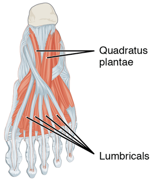
Quadratus plantae makes part of the 20 individual foot muscles. It is situated in the second layer of muscles at the sole of the foot. | Quadratus plantae makes part of the 20 individual foot muscles. It is situated in the second layer of muscles at the sole of the foot.<ref name=":0">Moore KL, Dalley AF, Agur AMR. Clinial oriented anatomy. Philadelphia: Wolters Kluwer, 2010.</ref> | ||
[[File:Intrinsicfoot mm.png|none|thumb]] | |||
=== Origin === | === Origin === | ||
Medial surface and lateral margin of the plantar surface of the [[calcaneus]]. | Medial surface and lateral margin of the plantar surface of the [[calcaneus]].<ref name=":0" /> | ||
=== Insertion === | === Insertion === | ||
Posterolateral margin of tendon of [[flexor digitorum longus]]. | Posterolateral margin of tendon of [[flexor digitorum longus]].<ref name=":0" /> | ||
=== Nerve === | === Nerve === | ||
Lateral plantar nerve (S2, S3). | Lateral plantar nerve (S2, S3).<ref name=":0" /> | ||
=== Artery === | === Artery === | ||
Lateral plantar artery. | Lateral plantar artery.<ref name=":0" /> | ||
== Function == | == Function == | ||
The muscles of the foot are arranged in compartments and layers, but function together to support the foot during stance phase and maintaining the arch of the foot. Quadratus plantae mainly functions by assisting [[flexor digitorum longus]] with flexion of the lateral 4 digits of the [[Foot Anatomy|foot]]. | The muscles of the foot are arranged in compartments and layers, but function together to support the foot during stance phase and maintaining the arch of the foot. Quadratus plantae mainly functions by assisting [[flexor digitorum longus]] with flexion of the lateral 4 digits of the [[Foot Anatomy|foot]].<ref name=":0" /> | ||
== Clinical relevance == | == Clinical relevance == | ||
Quadratus plantae increase the stability of the foot during the stance phase of gait to resist toe extension. It is thus an important foot muscle to consider in the [[Gait Cycle|gait pattern]] and with gait retraining after foot injuries. | |||
Quadratus plantae contractures can lead the clawing of 2nd to 5th toes, often following a [[Calcaneal Fractures|calcaneus fracture]].<ref>Sooriakumaran P, Sivananthan S. [https://pdfs.semanticscholar.org/f051/1368acfdf227be873672246f474f5227620f.pdf Why does man have a quadratus plantae? A review of its comparative anatomy.] Croatian medical journal 2005;46(1).</ref> | |||
The quadratus plantae lies deep within the posterior compartment of the hindfoot, with a communication to the posterior compartment of the leg through the retinaculum behind the medial malleolus, following the neurovascular and tendinous structures. Contracture of the quadratus plantae within this calcaneal compartment results in clawing of the lesser toes (digits two to five) as a late sequela of calcaneal fractures (11). The muscle is also subject to necrosis from untreated central plantar space abscesses in the diabetic foot. The quadratus plantae has also been implicated in the pathogenesis of heel pain by a number of authors (22). The lateral plantar nerve is of mixed type, consisting of sensory fibers for the calcaneal periosteum, plantar ligament, and medial head of quadratus plantae, and motor fibers to quadratus plantae. This means that when heel pain is initiated due to the entrapment of the lateral plantar nerve between the two heads, pain is felt over the calcaneus and the quadratus plantae muscle function is impaired (22). In a study concerning the lateral plantar nerve and heel pain, it was found that in fetal feet the first branch of this nerve penetrates the insertion of quadratus plantae. However, in adult feet this nerve always sends fibers to the periosteum around the medial process of the calcaneal tuberosity and long plantar ligament (22). Thus, it can be deduced that there may be redistribution in the nerve supply to this muscle during the development. In a case study by Murphy (23), repeated myofascial therapy to quadratus plantae over a 4-month period, in combination with manipulation therapy to the ankle and intertarsal joints, brought dramatic improvement in a patient’s symptoms and signs of diabetic polyneuropathy. From this study, it has been suggested that joint and/or myofascial dysfunction may be involved in the susceptibility to this condition and thus treatment may improve the neuropathy. The author proposes mechanisms by which this relationship may exist, including impaired afferent stimulation from the intertarsal joint receptors due to the loss of joint play and disturbed axoplasmic flow along the nerves affected by the neuropathy. Conclusions Phylogenetic evidence for the presence of quadratus plantae in humans is not found due to its abnormal development and morphology. A doubled-headed quadratus plantae is unique to man (6). The presence of quadratus plantae is subject to wide variation in man, and the human muscle is a composite structure where the lateral head is homologous to that in other mammals and the medial head is unique to man. Its role in realigning the pull generated by flexor digitorum longus can be supported by its site of insertion and position of the ankle joint but this effect is not nearly as important as had previously been advocated. Its role in suppressing inversion of the subtalar joint seems most plausible when the evolutionary standpoint is taken, yet electromyographic studies do not support this. Reeser (12) concluded that the main role of the quadratus plantae, together with the flexor abductor brevis, was to support flexor digitorum longus, and it had little, if any, role in eversion. Studies by Hicks (20,24) have suggested that the plantar aponeurosis is most important for active flexion of the toes. This reverse windlass mechanism postulates that flexion of the toes can be achieved without contraction of flexor digitorum longus, making the role of quadratus plantae as a support of flexor digitorum longus seem even less important. In summary, there is much conflicting evidence regarding the role of quadratus plantae. Further work is required to resolve this issu | |||
== Assessment == | == Assessment == | ||
* Palpation | |||
== Treatment == | == Treatment == | ||
| Line 36: | Line 43: | ||
* [[Foot Anatomy|Foot anatomy]] | * [[Foot Anatomy|Foot anatomy]] | ||
* [[Ankle and Foot|Ankle and foot]] | * [[Ankle and Foot|Ankle and foot]] | ||
== References == | == References == | ||
Revision as of 22:13, 30 March 2020
Original Editor - Leana Louw
Top Contributors - Leana Louw, Patti Cavaleri and Kim Jackson
This article or area is currently under construction and may only be partially complete. Please come back soon to see the finished work! (30/03/2020)
Description[edit | edit source]
Quadratus plantae makes part of the 20 individual foot muscles. It is situated in the second layer of muscles at the sole of the foot.[1]
Origin[edit | edit source]
Medial surface and lateral margin of the plantar surface of the calcaneus.[1]
Insertion[edit | edit source]
Posterolateral margin of tendon of flexor digitorum longus.[1]
Nerve[edit | edit source]
Lateral plantar nerve (S2, S3).[1]
Artery[edit | edit source]
Lateral plantar artery.[1]
Function[edit | edit source]
The muscles of the foot are arranged in compartments and layers, but function together to support the foot during stance phase and maintaining the arch of the foot. Quadratus plantae mainly functions by assisting flexor digitorum longus with flexion of the lateral 4 digits of the foot.[1]
Clinical relevance[edit | edit source]
Quadratus plantae increase the stability of the foot during the stance phase of gait to resist toe extension. It is thus an important foot muscle to consider in the gait pattern and with gait retraining after foot injuries.
Quadratus plantae contractures can lead the clawing of 2nd to 5th toes, often following a calcaneus fracture.[2]
The quadratus plantae lies deep within the posterior compartment of the hindfoot, with a communication to the posterior compartment of the leg through the retinaculum behind the medial malleolus, following the neurovascular and tendinous structures. Contracture of the quadratus plantae within this calcaneal compartment results in clawing of the lesser toes (digits two to five) as a late sequela of calcaneal fractures (11). The muscle is also subject to necrosis from untreated central plantar space abscesses in the diabetic foot. The quadratus plantae has also been implicated in the pathogenesis of heel pain by a number of authors (22). The lateral plantar nerve is of mixed type, consisting of sensory fibers for the calcaneal periosteum, plantar ligament, and medial head of quadratus plantae, and motor fibers to quadratus plantae. This means that when heel pain is initiated due to the entrapment of the lateral plantar nerve between the two heads, pain is felt over the calcaneus and the quadratus plantae muscle function is impaired (22). In a study concerning the lateral plantar nerve and heel pain, it was found that in fetal feet the first branch of this nerve penetrates the insertion of quadratus plantae. However, in adult feet this nerve always sends fibers to the periosteum around the medial process of the calcaneal tuberosity and long plantar ligament (22). Thus, it can be deduced that there may be redistribution in the nerve supply to this muscle during the development. In a case study by Murphy (23), repeated myofascial therapy to quadratus plantae over a 4-month period, in combination with manipulation therapy to the ankle and intertarsal joints, brought dramatic improvement in a patient’s symptoms and signs of diabetic polyneuropathy. From this study, it has been suggested that joint and/or myofascial dysfunction may be involved in the susceptibility to this condition and thus treatment may improve the neuropathy. The author proposes mechanisms by which this relationship may exist, including impaired afferent stimulation from the intertarsal joint receptors due to the loss of joint play and disturbed axoplasmic flow along the nerves affected by the neuropathy. Conclusions Phylogenetic evidence for the presence of quadratus plantae in humans is not found due to its abnormal development and morphology. A doubled-headed quadratus plantae is unique to man (6). The presence of quadratus plantae is subject to wide variation in man, and the human muscle is a composite structure where the lateral head is homologous to that in other mammals and the medial head is unique to man. Its role in realigning the pull generated by flexor digitorum longus can be supported by its site of insertion and position of the ankle joint but this effect is not nearly as important as had previously been advocated. Its role in suppressing inversion of the subtalar joint seems most plausible when the evolutionary standpoint is taken, yet electromyographic studies do not support this. Reeser (12) concluded that the main role of the quadratus plantae, together with the flexor abductor brevis, was to support flexor digitorum longus, and it had little, if any, role in eversion. Studies by Hicks (20,24) have suggested that the plantar aponeurosis is most important for active flexion of the toes. This reverse windlass mechanism postulates that flexion of the toes can be achieved without contraction of flexor digitorum longus, making the role of quadratus plantae as a support of flexor digitorum longus seem even less important. In summary, there is much conflicting evidence regarding the role of quadratus plantae. Further work is required to resolve this issu
Assessment[edit | edit source]
- Palpation


