Quadriceps Muscle Strain: Difference between revisions
No edit summary |
No edit summary |
||
| (168 intermediate revisions by 21 users not shown) | |||
| Line 1: | Line 1: | ||
<div class="editorbox"> | |||
'''Original Editors ''' - [[User:Maxime Tuerlinckx|Maxime Tuerlinckx]] | '''>Original Editors ''' - [[User:Maxime Tuerlinckx|Maxime Tuerlinckx]] | ||
''' | '''Top Contributors''' - {{Special:Contributors/{{FULLPAGENAME}}}}<br> | ||
</div> | </div> | ||
== | == Introduction == | ||
[[File:Quadriceps femoris muscle - Kenhub.png|alt=Quadriceps femoris muscle (highlighted in green) - anterior view|400x400px|Quadriceps femoris muscle (green) |thumb]] | |||
A [[Quadriceps Muscle|quadriceps]] muscle strain is an acute tearing injury of the quadriceps.This injury is usually due to an acute [[Stretching|stretch]] of the [[muscle]], often at the same time of a forceful contraction or repetitive functional overloading. <ref name=":0">Kary JM. [https://www.ncbi.nlm.nih.gov/pmc/articles/PMC2941577/ Diagnosis and management of quadriceps strains and contusions]. Current reviews in musculoskeletal medicine. 2010 Oct 1;3(1-4):26-31.</ref> | |||
Injury to the quadriceps muscle group can be painful and debilitating. [[Muscle Strain|Strains]] and [[Quadriceps Muscle Contusion|contusions of the quadriceps]] are common in athletics and result in lost time from training and competition.<ref name=":2">Kary JM. Diagnosis and management of quadriceps strains and contusions. Current reviews in musculoskeletal medicine. 2010 Oct;3(1):26-31.Available;https://www.ncbi.nlm.nih.gov/pmc/articles/PMC2941577/ (accessed 4.12.2022)</ref> | |||
'''Clinically Relevant Anatomy:''' The [[Quadriceps Muscle|Quadriceps femoris]] is a hip flexor and a knee extensor. It is located in the anterior compartment of the thigh. See link for more. This muscle is composed of 4 sub components: [https://physio-pedia.com/Rectus_Femoris Rectus femoris (RF)]; [[Vastus Lateralis|Vastus lateralis]];[[Vastus Medialis|Vastus medialis]]; [[Vastus Intermedius|Vastus intermedius]]. | |||
== Epidemiology == | |||
[[File:Sprinting.jpeg|thumb|350x350px|Sprinting: risk factor for RF strain]] | |||
Quadriceps injuries are common injuries in athletes with the muscle prone to muscle strains in situations requiring explosive movements as seen in certain sports. | |||
== | * Rectus femoris muscle is the most commonly involved in a quadriceps injury, factors predisposing RF include crossing two joints, a high percentage of Type II fibers, and having a complex musculotendinous architecture<ref name=":2" /> | ||
* Vastus muscle injury the least common, and the vastus intermedius muscle is most commonly affected by contusions. | |||
* Quadriceps tendon rupture is uncommon debilitating diagnosis, occurring more often in older men.<ref name=":3">Radiopedia Quadriceps injury Available:https://radiopaedia.org/articles/quadriceps-injury (accessed 4.12.2022)</ref> | |||
== Etiology == | |||
[[File:Hurdles overstretched RF.jpeg|thumb|Hurdles possible overstretched RF]] | |||
Acute strain injuries of the quadriceps commonly occur: | |||
* In sports that regularly require sudden forceful eccentric contraction of the quadriceps during regulation of knee flexion and hip extension eg soccer, rugby, and football. | |||
* When high forces occur across the muscle–tendon units with eccentric contraction. | |||
* When excessive passive stretching or activation of a maximally stretched muscle occurs. | |||
* When a muscle is fatigued<ref name=":2" /> | |||
There are generally three mechanisms of injury for a quadriceps strain:<br>1. Sudden deceleration of the leg (e.g. kicking), <br>2. violent contraction of the quadriceps (sprinting) and <br>3. rapid deceleration of an overstretched muscle (by quickly change of direction). | |||
== Classification of Quadriceps Strains == | |||
Below provided is an outline of a clinical grading system for muscle strains. Factoring in pain, loss of strength, and physical exam findings in a grading system helps provide guidance for treatment, rehabilitation, and eventual return to play.<ref name=":2" /> | |||
# '''Grade I''' (mild) strains affect only a limited number of fibers in the muscle. There is no decrease in strength and there is a fully active and passive range of motion. Pain and tenderness are often delayed to the next day. | |||
# '''Grade II''' (moderate) strains have nearly half of muscle fibers torn. Acute and significant pain is accompanied by swelling and a minor decrease in muscle strength. | |||
# '''Grade III''' (severe) strains represent the complete rupture of the muscle. This means either the tendon is separated from the muscle belly or the muscle belly is actually torn in 2 parts. Severe swelling and pain and a complete loss of function are characteristic of this type of strain.<ref>[[Muscle Strain]]</ref> | |||
Muscle injuries also be broadly classified as either acute or chronic injuries. | |||
# '''Acute injuries''': are usually the result of a single traumatic event and cause a macro-trauma to the muscle. There is an obvious link between the cause and noticeable symptoms.They mostly occur in contact sports such as rugby, soccer and basketball because of their dynamic and high collision nature.<ref name=":1">Best TM. [https://www.ncbi.nlm.nih.gov/pubmed/9209819 Soft-tissue injuries and muscle tears]. Clinics in sports medicine. 1997 Jul 1;16(3):419-34.</ref><ref>Beiner JM, Jokl P. [https://www.ncbi.nlm.nih.gov/pubmed/11476532 Muscle contusion injuries: current treatment options.] JAAOS-Journal of the American Academy of Orthopaedic Surgeons. 2001 Jul 1;9(4):227-37.</ref> | |||
# '''Overuse:('''chronic or exercise-induced injuries)are subtler and usually occur over a longer period of time.They result from repetitive micro-trauma to the muscle. Diagnosing is more challenging since there is a less obvious link between the cause of the injury and the symptoms.<ref name=":1" /> | |||
==== Grades of quadriceps strain ==== | |||
# <u>'''Grade 1 symptoms:'''</u> Symptoms of a grade 1 quadriceps strain are not always serious enough to stop training at the time of injury. A twinge may be felt in the thigh and a general feeling of tightness.The athlete may feel mild discomfort on walking and running might be difficult.There is unlikely to be swelling. A lump or area of spasm at the site of injury may be felt. | |||
# <u>'''Grade 2 symptoms:'''</u> The athlete may feel a sudden sharp pain when running, jumping or kicking and be unable to play on.Pain will make walking difficult and swelling or mild bruising may be noticed.The pain would be felt when pressing in on the suspected location of the quad muscle tear.Straightening the knee against resistance is likely to cause pain and the injured athlete will be unable to fully bend the knee. | |||
# <u>'''Grade 3 symptoms:'''</u> Symptoms consist of a severe,sudden pain in the front of the thigh.The patient will be unable to walk without the aid of crutches.Bad swelling will appear immediately and significant bruising within 24 hours.A static muscle contraction will be painful and is likely to produce a bulge in the muscle.The patient can expect to be out of competition for 6 to 12 weeks.<ref>Thigh Strain http://www.sportsinjuryclinic.net/sport-injuries/thigh-pain/quadriceps-strain (Last accessed 22 july 2018) | |||
</ref> | |||
== Differential Diagnosis == | |||
*Soft tissue tumours/ bone [[Oncology|tumours]] | |||
*[[Myositis Ossificans of the Quadriceps|Myositis ossificans]]<ref name=":3" /> | |||
== Diagnosis == | |||
Most muscle injuries can be elucidated by a thorough history and physical examination.<ref name="Tero">Tero AH Järvinen, Markku Järvinen, Hannu Kalimo; Regeneration of injured skeletal muscle after the injury; Muscles, Ligaments and Tendons Journal 2013; 3 (4): 337-345 (2A)</ref> | |||
# Quadriceps muscle strains have a variable clinical history ranging from a sharp thigh pain and/or hip pain associated with movements to vague aches or thigh enlargement and variably associated strength deficit. Tenderness upon direct palpation is a typical finding and pain can usually be triggered by stretching and resisted muscle activity. | |||
# [[Medical Imaging|Imaging]] and in particular [[Ultrasound Scans|ultrasound]] and [[MRI Scans|MRI]] can provide information regarding the extent, type and prognosis of the muscle injury.<ref name=":3" /> | |||
== Examination == | |||
After obtaining a thorough [[Subjective Assessment|history]], a careful examination should ensue including observation, palpation, strength testing, and evaluation of motion. | |||
* '''Observation:''' The therapist will have a close look at the injured area, observing for swelling and bruising in particular.They should also observe the patient in standing and walking, looking for postural abnormalities. They may present with an obvious deformity such as a bulge or defect in the muscle belly. | |||
* '''Palpation:''' Palpation of the quadriceps muscle should occur along the entire length of the muscles and the aponeuroses.This is required to identify swelling, thickening, tenderness, defects and masses if present. Acute compartment syndrome should be considered if there is tenseness of the fascial envelope surrounding the compartment and pain out of proportion to the clinical situation. | |||
* '''Strength testing:''' of the quadriceps should include resistance of knee extension and hip flexion. Adequate strength testing of the rectus femoris must include resisted knee extension with the hip flexed and extended. Practically, this is best accomplished by evaluating the patient in both a sitting and prone-lying position. The prone-lying position also allows for optimum assessment of quadriceps motion and flexibility. Pain is typically felt by the patient with resisted muscle activation, passive stretching, and direct palpation over the muscle strain. | |||
* '''Assessing tenderness:'''any palpable defect and strength at the onset of muscle injury will determine grading of the injury and provide direction for further diagnostic testing and treatment.  | |||
==='''Outcome Measures'''=== | |||
* [[Muscle Strength Testing]] | |||
* [[Range of Motion|ROM]]. | |||
*Voluntary activation by superimposing percutaneous electrical stimulation on to an isometric quadriceps. When the muscle is fully activated, the electrical stimulation does not generate additional force above the voluntary contraction. | |||
== Medical Management == | |||
Management of quadriceps injuries depends on the type and location of the injury. A complete quadriceps tendon tear with loss of the extensor mechanism requires surgery and reattachment of the quadriceps tendon. See [[Muscle Rupture Surgery]].<ref name=":3" /> All other quadriceps injuries can be managed conservatively using the accepted method to treat muscle strain injuries in the initial period, being the [[RICE]] (rest, ice, compression and limb elevation) principle or the [[POLICE Principle|POLICE]] principle. | |||
== Physical Therapy Management == | |||
Acute Phase: An initial resting period serves as a measure to prevent further progression of the injury and a more severe strain injury might require an initial use of crutches. Limb elevation and intermittent application of ice and compression are aiming to decrease blood flow and increased amounts of interstitial fluid accumulation. Ice application also serves pain control and can be supplemented by non-steroidal anti-inflammatory drugs (NSAIDs) in the initial period. <span style="line-height: 1.5em;">The use of [[NSAIDs|NSAID]]'s is still controversial, their benefit, cost and potential adverse effects may be taken into consideration. If used, it should be during the inflammatory period (48h-72h) </span><ref name="p1">Almekinders LC. Anti-inflammatory treatment of muscular injuries in sport. An update of recent studies. Sports Med. Dec 1999;28(6):383-8.</ref>. | |||
Knee Positioning: When a quadriceps muscle strain occurs during a competition or training, it is important to react immediately. In the 10 minutes following the trauma one needs to put the knee of the affected leg immediately in 120° of flexion'''.<ref name="p1" /><ref name="p2">Michael A Herbenick, MD; Michael S Omori, MD; Paul Fenton, MD. Contusions, 2009 (A)</ref> '''This avoids potential muscle spasms, reduces the hemorrhage and minimizes the risk of developing [http://www.physio-pedia.com/Myositis_Ossificans_of_the_Quadriceps myositis ossificans]<ref name="p2" />.Practically, this can be done by placing the patient in a hinged knee brace at 120° of knee flexion or using elastic compression wrap to maintain this position of flexion.If the knee is left in extension the healing process will be slower and more painful because the quadriceps will start to heal in a shortened position.<ref name="p2" /> | |||
=== Active Phase of Management === | |||
[[File:Quadriceps Stretch.png|alt=|thumb|Quadriceps Stretch]][[File:Hip Flexor Stretch.png|thumb|alt=|Hip Flexor Stretc]]The acute phase of treatment is subsequently followed by an active phase of management once the injured leg is recovering well. This phase usually begins approximately 3–5 days after the initial injury depending on its severity. Stretching, strengthening, range of motion, maintenance of aerobic fitness, proprioceptive exercises, and functional training are the primary components of this phase.<ref name=":02">Kary JM. [https://www.ncbi.nlm.nih.gov/pmc/articles/PMC2941577/ Diagnosis and management of quadriceps strains and contusions]. Current reviews in musculoskeletal medicine. 2010 Oct 1;3(1-4):26-31.</ref>. | |||
# '''<u>Stretching</u>''' : Stretching should be done carefully and always to the point of discomfort, but not pain. Various techniques can be utilized including passive, active–passive, dynamic, and proprioceptive neuromuscular facilitation stretching. Generally, ballistic stretching is discouraged due to the risk of re-tearing muscle fibers. If it is pain free , stretch the quad muscles. | |||
'''Static quad stretch''' :This can be performed in either standing, or laying on your front. Pull the foot of the injured leg towards your buttock until you can feel a gentle stretch on the front of the thigh. To increase the stretch, tilt your hips backwards. Hold for 20-30 seconds and repeat 3 times. Do this at least 3 times a day. | |||
'''Hip flexor stretch:'''This stretch will focus on the rectus femoris and Iliopsoas muscles. Kneel with one knee on the floor and the other foot out in front with the knee bent. Push your hips forwards and keep the back upright. You should feel a stretch at the front of the hip and top of the thigh.Hold for 20-30 seconds, repeat 3 times, at least 3 times a day. | |||
'''2.<u>Strengthening exercises:</u>''' | |||
The aim of strengthening exercises is to gradually increase the load that is put through a muscle. Strengthening exercises can start as early as day 5 as long as they are low-level and must be done pain-free.Isometric or static exercises are advised first and then progress to dynamic exercises with resistance band and finishing with sports specific running and sprint drills. scientific evidence is lacking on the consensus of treatment principles of muscle injuries <ref name=":0" /> | |||
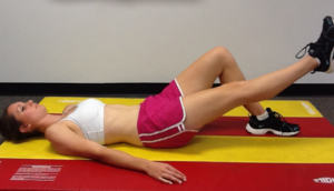
* '''Isometrics''' : Initial isometrics with quadriceps contractions done with the knee fully extended and in different positions at 20 degree increments as knee flexion improves May discontinue isometrics when patient can sit comfortable. | |||
<div class="row"> | |||
<div class="col-md-6"> {{#ev:youtube|9EhHFemc8WQ}} <div class="text-right"><ref>Knee exercise for knee pain - Isometric quads. Available from https://www.youtube.com/watch?v=9EhHFemc8WQ . (last accessed 5 August 2018)</ref></div></div> | |||
<div class="col-md-6"> {{#ev:youtube|nPvgiRjsEIs}} <div class="text-right"><ref>Quad exercises - isometric quads prone.Available from https://www.youtube.com/watch?v=nPvgiRjsEIs .(last accessed 5 August 2018)</ref></div></div> | |||
*[[File:Straight leg raise.png|frameless|alt=|right]]'''Straight leg raises''' : Sit flat on the floor with the legs straight out in front of you. Raise one leg off the floor keeping the knee straight. Hold for 3 to 5 seconds before lowering back to the ground. Repeat 10 to 20 times. This exercise can be done daily. Progress the exercise by increasing the length of hold and the number of reps. | |||
* '''Isotonics''' : Once terminal knee extensions are done properly without extensor lag, freeweights are added to the SLRs and terminal knee extensions . Begin with the lightest free weight that patient can lift; three sets of 10 repetitions up to three times per day.Increase weight by no more than 2-3 pounds at any given time and increase no sooner than every two consecutive work days. | |||
* '''Wall squats :''' From your starting position, slowly lower your body down and hold for time. As you improve, lengthen the amount of time you hold the wall squat. Be sure to keep your pelvis, back, and head against the wall. Keep the movement pain free.(A variation to increase activation of the VMO would be to squeeze a ball between your knees as you perform the exercise.Typically the ball would be about 12 inches in diameter.)Perform 3 sets of 15-20 seconds holds once per day. | |||
{{#ev:youtube|MMV3v4ap4ro}}<ref>Wall Sit.Passion4Profession. Available from https://www.youtube.com/watch?v=MMV3v4ap4ro .(last accessed 5 August 2018)</ref> | |||
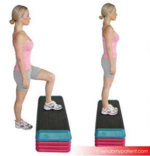
*[[File:Stepping.gif|frameless|alt=|right]]'''Step ups''' : Start with a box height that is comfortable for you to step up on. Be sure to keep your knee in alignment with your second toe. Step up and keep your pelvis level and your knee in alignment. Be sure to engage the buttocks muscles and fully lock out the knee. Return slowly back down to the ground. The focus should be on the slow eccentric (lowering) back to the ground for 1 second up and 3 seconds down.Perform 2 sets of 15-20 repetitions once per day. Technique: Stand facing a step. Place your affected leg up on the step. Step up bringing your other leg onto the step and then step back down to the start position using the same leg. Make sure your knee travels forwards over your toes during this exercise. Your affected leg will stay on the step throughout this exercise. | |||
== Rehabilitation Protocol Example == | |||
==<ref>Rehabilitation of Quadriceps Strain http://orthodoc.aaos.org/Hartman/Quadriceps%20Strain.pdf (accessed on 9 August 2018)</ref>== | |||
Acute Phase week 1-2 | |||
= | {| class="wikitable" | ||
!Goals: | |||
|Diminish pain and inflammation | |||
Gradually improve flexibility and ROM | |||
Retard muscular atrophy and strength loss | |||
= | Enhance healing of muscular strain | ||
|- | |||
!precautions | |||
|Avoid excessive active or passive lengthening of quadriceps | |||
|- | |||
!Rehab | |||
| | |||
* RICE–Rest,Cryotherapy, compression wrap, and elevation | |||
* Use of crutches initially to facilitate rest and immobilization of the quadriceps | |||
* NSAIDS | |||
* Soft tissue mobs/IASTM | |||
* Pulsed ultrasound (Duty cycle 50%, 1 MHz, 1.2 W/cm2) | |||
* Conventional TENS | |||
* Ankle pumps, isometric quadriceps sets, hamstring sets, glut sets | |||
|} | |||
* '''PHASE 2 :SUBACUTE PHASE (WEEK 3 - 12)''' | |||
{| class="wikitable" | |||
!Goals | |||
|Regain pain-free quadriceps strength, progressing through full ROM | |||
Develop neuromuscular control of trunk and pelvis with progressive increase in movement and speed preparing for functional movements | |||
|- | |||
!precautions | |||
|Avoid end-range lengthening of quadriceps if painful | |||
|- | |||
!Rehab | |||
| | |||
* Cryotherapy | |||
* NSAIDS | |||
* Electrical stimulation | |||
* Initial isometrics with quadriceps contractions done with the knee fully extended and in different positions at 20 degree increments as knee flexion improves | |||
* May discontinue isometrics when can sit comfortably, perform straight leg raises at 0 degrees, 20 degrees, and 40 degrees | |||
* Isotonics–begin with the lightest free weight that athlete can lift; three sets of 10 repetitions up to three times per day | |||
* Terminal knee extensions instituted at 20 degree increments as comfort and knee flexion allow | |||
* Once terminal knee extensions are done properly without extensor lag, free weights are added to the SLRs and terminal knee extensions | |||
* Increase weight by no more than 2-3 pounds at any given time and increase no sooner than every two consecutive work days | |||
* As athlete approaches his or her maximum weight, somewhere around 15-20pounds, isokinetic exercises are tried | |||
* Conditioning via upper body workouts, swimming, treadmill walking | |||
* Biking when knee ROM greater than 100 degrees of flexion | |||
|} | |||
* '''Phase 3 ( week 12+)''' | |||
{| class="wikitable" | |||
!Goals | |||
|Symptom free during all activities | |||
Normal concentric and eccentric strength through full ROM and speed | |||
Improve neuromuscular control of trunk and pelvis | |||
Integrate postural control into sport-specific movements | |||
|- | |||
!Precautions | |||
|Train within symptoms free intensity | |||
|- | |||
!Rehab | |||
| | |||
* Ice – Post exercise – as needed | |||
* Treadmill moderate to high intensity as tolerated | |||
* Isokinetic eccentric training at end ROM (in hyperflexion) | |||
* STM/IASTM | |||
* Plyometric jump training | |||
* 5-10 yard accelerations/decelarations | |||
* Single-limb balance windmill touches with weight on unstable surface | |||
* Sport-specific drills that incorporate postural control and progressive speed | |||
|- | |||
!Eccentric protocol | |||
| | |||
* Include higher velocity eccentric Ex that include plyometric and sports specific activities | |||
* Examples: include squat jumps, split jumps, bounding and depth jumps, single leg bounding, backward skips, lateral hops, lateral bounding, zigzag hops, bounding, plyometric box jumps, eccentric backward steps, eccentric lunge drops, eccentric forward pulls, single and double leg deadlifts, and split stance deadlift (good morning Ex) | |||
|} | |||
=== Return to Sports Criteria === | |||
< | * Full strength without pain in the lengthened state testing position | ||
* Full ROM without pain | |||
</ | * Replication of sport specific movements at competition speed without symptoms. | ||
* Isokinetic strength testing should be performed under both concentric and eccentric action conditions. | |||
* Attain 120° of knee flexion with hip extended<ref>Kary, Joel M. "Diagnosis and management of quadriceps strains and contusions." Current reviews in musculoskeletal medicine 3.1-4 (2010): 26-31. </ref> | |||
== Clinical Bottom Line == | |||
<br>Acute strain injuries of the quadriceps commonly occur in athletic competitions such as soccer, rugby, and football. These sports regularly require sudden forceful eccentric contraction of the quadriceps during regulation of knee flexion and hip extension. Higher forces across the muscle–tendon units with eccentric contraction can lead to strain injury. Excessive passive stretching or activation of a maximally stretched muscle can also cause strains. Of the quadriceps muscles, the rectus femoris is most frequently strained. Several factors predispose this muscle and others to more frequent strain injury. These include muscles crossing two joints, those with a high percentage of Type II fibers, and muscles with complex musculotendinous architecture. Muscle fatigue has also been shown to play a role in acute muscle injury.<br>Therapy is bases on 3 principles: <br>1. POLICE or RICE <br>2. Knee mobilisation <br>3. Training of the quadriceps functions | |||
<references /> | == References == | ||
<references /> | |||
[[Category:Vrije_Universiteit_Brussel_Project | [[Category:Injury]] | ||
[[Category:Knee_Injuries]] | |||
[[Category:Knee]] | |||
[[Category:Conditions]] | |||
[[Category:Knee - Conditions]] | |||
[[Category:Muscles]] | |||
[[Category:Musculoskeletal/Orthopaedics|Orthopaedics]] | |||
[[Category:Vrije_Universiteit_Brussel_Project]] | |||
[[Category:Sports_Injuries]] | |||
[[Category:Sports Medicine]] | |||
[[Category:Primary Contact]] | |||
Latest revision as of 02:18, 5 December 2022
>Original Editors - Maxime Tuerlinckx
Top Contributors - Mandeepa Kumawat, Maxime Tuerlinckx, Carlos Areia, Lucinda hampton, Shanna Blyckaerts, Admin, Shaimaa Eldib, Wanda van Niekerk, Jelle Van Hemelryck, Kim Jackson, Rachael Lowe, Frederik Töpke, WikiSysop, Joao Costa, Claire Knott, Rucha Gadgil, Evan Thomas, Scott Buxton, Naomi O'Reilly, Daphne Jackson, Fasuba Ayobami and 127.0.0.1
Introduction[edit | edit source]

A quadriceps muscle strain is an acute tearing injury of the quadriceps.This injury is usually due to an acute stretch of the muscle, often at the same time of a forceful contraction or repetitive functional overloading. [1]
Injury to the quadriceps muscle group can be painful and debilitating. Strains and contusions of the quadriceps are common in athletics and result in lost time from training and competition.[2]
Clinically Relevant Anatomy: The Quadriceps femoris is a hip flexor and a knee extensor. It is located in the anterior compartment of the thigh. See link for more. This muscle is composed of 4 sub components: Rectus femoris (RF); Vastus lateralis;Vastus medialis; Vastus intermedius.
Epidemiology[edit | edit source]

Quadriceps injuries are common injuries in athletes with the muscle prone to muscle strains in situations requiring explosive movements as seen in certain sports.
- Rectus femoris muscle is the most commonly involved in a quadriceps injury, factors predisposing RF include crossing two joints, a high percentage of Type II fibers, and having a complex musculotendinous architecture[2]
- Vastus muscle injury the least common, and the vastus intermedius muscle is most commonly affected by contusions.
- Quadriceps tendon rupture is uncommon debilitating diagnosis, occurring more often in older men.[3]
Etiology[edit | edit source]

Acute strain injuries of the quadriceps commonly occur:
- In sports that regularly require sudden forceful eccentric contraction of the quadriceps during regulation of knee flexion and hip extension eg soccer, rugby, and football.
- When high forces occur across the muscle–tendon units with eccentric contraction.
- When excessive passive stretching or activation of a maximally stretched muscle occurs.
- When a muscle is fatigued[2]
There are generally three mechanisms of injury for a quadriceps strain:
1. Sudden deceleration of the leg (e.g. kicking),
2. violent contraction of the quadriceps (sprinting) and
3. rapid deceleration of an overstretched muscle (by quickly change of direction).
Classification of Quadriceps Strains[edit | edit source]
Below provided is an outline of a clinical grading system for muscle strains. Factoring in pain, loss of strength, and physical exam findings in a grading system helps provide guidance for treatment, rehabilitation, and eventual return to play.[2]
- Grade I (mild) strains affect only a limited number of fibers in the muscle. There is no decrease in strength and there is a fully active and passive range of motion. Pain and tenderness are often delayed to the next day.
- Grade II (moderate) strains have nearly half of muscle fibers torn. Acute and significant pain is accompanied by swelling and a minor decrease in muscle strength.
- Grade III (severe) strains represent the complete rupture of the muscle. This means either the tendon is separated from the muscle belly or the muscle belly is actually torn in 2 parts. Severe swelling and pain and a complete loss of function are characteristic of this type of strain.[4]
Muscle injuries also be broadly classified as either acute or chronic injuries.
- Acute injuries: are usually the result of a single traumatic event and cause a macro-trauma to the muscle. There is an obvious link between the cause and noticeable symptoms.They mostly occur in contact sports such as rugby, soccer and basketball because of their dynamic and high collision nature.[5][6]
- Overuse:(chronic or exercise-induced injuries)are subtler and usually occur over a longer period of time.They result from repetitive micro-trauma to the muscle. Diagnosing is more challenging since there is a less obvious link between the cause of the injury and the symptoms.[5]
Grades of quadriceps strain[edit | edit source]
- Grade 1 symptoms: Symptoms of a grade 1 quadriceps strain are not always serious enough to stop training at the time of injury. A twinge may be felt in the thigh and a general feeling of tightness.The athlete may feel mild discomfort on walking and running might be difficult.There is unlikely to be swelling. A lump or area of spasm at the site of injury may be felt.
- Grade 2 symptoms: The athlete may feel a sudden sharp pain when running, jumping or kicking and be unable to play on.Pain will make walking difficult and swelling or mild bruising may be noticed.The pain would be felt when pressing in on the suspected location of the quad muscle tear.Straightening the knee against resistance is likely to cause pain and the injured athlete will be unable to fully bend the knee.
- Grade 3 symptoms: Symptoms consist of a severe,sudden pain in the front of the thigh.The patient will be unable to walk without the aid of crutches.Bad swelling will appear immediately and significant bruising within 24 hours.A static muscle contraction will be painful and is likely to produce a bulge in the muscle.The patient can expect to be out of competition for 6 to 12 weeks.[7]
Differential Diagnosis[edit | edit source]
- Soft tissue tumours/ bone tumours
- Myositis ossificans[3]
Diagnosis[edit | edit source]
Most muscle injuries can be elucidated by a thorough history and physical examination.[8]
- Quadriceps muscle strains have a variable clinical history ranging from a sharp thigh pain and/or hip pain associated with movements to vague aches or thigh enlargement and variably associated strength deficit. Tenderness upon direct palpation is a typical finding and pain can usually be triggered by stretching and resisted muscle activity.
- Imaging and in particular ultrasound and MRI can provide information regarding the extent, type and prognosis of the muscle injury.[3]
Examination[edit | edit source]
After obtaining a thorough history, a careful examination should ensue including observation, palpation, strength testing, and evaluation of motion.
- Observation: The therapist will have a close look at the injured area, observing for swelling and bruising in particular.They should also observe the patient in standing and walking, looking for postural abnormalities. They may present with an obvious deformity such as a bulge or defect in the muscle belly.
- Palpation: Palpation of the quadriceps muscle should occur along the entire length of the muscles and the aponeuroses.This is required to identify swelling, thickening, tenderness, defects and masses if present. Acute compartment syndrome should be considered if there is tenseness of the fascial envelope surrounding the compartment and pain out of proportion to the clinical situation.
- Strength testing: of the quadriceps should include resistance of knee extension and hip flexion. Adequate strength testing of the rectus femoris must include resisted knee extension with the hip flexed and extended. Practically, this is best accomplished by evaluating the patient in both a sitting and prone-lying position. The prone-lying position also allows for optimum assessment of quadriceps motion and flexibility. Pain is typically felt by the patient with resisted muscle activation, passive stretching, and direct palpation over the muscle strain.
- Assessing tenderness:any palpable defect and strength at the onset of muscle injury will determine grading of the injury and provide direction for further diagnostic testing and treatment.
Outcome Measures[edit | edit source]
- Voluntary activation by superimposing percutaneous electrical stimulation on to an isometric quadriceps. When the muscle is fully activated, the electrical stimulation does not generate additional force above the voluntary contraction.
Medical Management[edit | edit source]
Management of quadriceps injuries depends on the type and location of the injury. A complete quadriceps tendon tear with loss of the extensor mechanism requires surgery and reattachment of the quadriceps tendon. See Muscle Rupture Surgery.[3] All other quadriceps injuries can be managed conservatively using the accepted method to treat muscle strain injuries in the initial period, being the RICE (rest, ice, compression and limb elevation) principle or the POLICE principle.
Physical Therapy Management[edit | edit source]
Acute Phase: An initial resting period serves as a measure to prevent further progression of the injury and a more severe strain injury might require an initial use of crutches. Limb elevation and intermittent application of ice and compression are aiming to decrease blood flow and increased amounts of interstitial fluid accumulation. Ice application also serves pain control and can be supplemented by non-steroidal anti-inflammatory drugs (NSAIDs) in the initial period. The use of NSAID's is still controversial, their benefit, cost and potential adverse effects may be taken into consideration. If used, it should be during the inflammatory period (48h-72h) [9].
Knee Positioning: When a quadriceps muscle strain occurs during a competition or training, it is important to react immediately. In the 10 minutes following the trauma one needs to put the knee of the affected leg immediately in 120° of flexion.[9][10] This avoids potential muscle spasms, reduces the hemorrhage and minimizes the risk of developing myositis ossificans[10].Practically, this can be done by placing the patient in a hinged knee brace at 120° of knee flexion or using elastic compression wrap to maintain this position of flexion.If the knee is left in extension the healing process will be slower and more painful because the quadriceps will start to heal in a shortened position.[10]
Active Phase of Management[edit | edit source]


The acute phase of treatment is subsequently followed by an active phase of management once the injured leg is recovering well. This phase usually begins approximately 3–5 days after the initial injury depending on its severity. Stretching, strengthening, range of motion, maintenance of aerobic fitness, proprioceptive exercises, and functional training are the primary components of this phase.[11].
- Stretching : Stretching should be done carefully and always to the point of discomfort, but not pain. Various techniques can be utilized including passive, active–passive, dynamic, and proprioceptive neuromuscular facilitation stretching. Generally, ballistic stretching is discouraged due to the risk of re-tearing muscle fibers. If it is pain free , stretch the quad muscles.
Static quad stretch :This can be performed in either standing, or laying on your front. Pull the foot of the injured leg towards your buttock until you can feel a gentle stretch on the front of the thigh. To increase the stretch, tilt your hips backwards. Hold for 20-30 seconds and repeat 3 times. Do this at least 3 times a day.
Hip flexor stretch:This stretch will focus on the rectus femoris and Iliopsoas muscles. Kneel with one knee on the floor and the other foot out in front with the knee bent. Push your hips forwards and keep the back upright. You should feel a stretch at the front of the hip and top of the thigh.Hold for 20-30 seconds, repeat 3 times, at least 3 times a day.
2.Strengthening exercises:
The aim of strengthening exercises is to gradually increase the load that is put through a muscle. Strengthening exercises can start as early as day 5 as long as they are low-level and must be done pain-free.Isometric or static exercises are advised first and then progress to dynamic exercises with resistance band and finishing with sports specific running and sprint drills. scientific evidence is lacking on the consensus of treatment principles of muscle injuries [1]
- Isometrics : Initial isometrics with quadriceps contractions done with the knee fully extended and in different positions at 20 degree increments as knee flexion improves May discontinue isometrics when patient can sit comfortable.
- Straight leg raises : Sit flat on the floor with the legs straight out in front of you. Raise one leg off the floor keeping the knee straight. Hold for 3 to 5 seconds before lowering back to the ground. Repeat 10 to 20 times. This exercise can be done daily. Progress the exercise by increasing the length of hold and the number of reps.

- Isotonics : Once terminal knee extensions are done properly without extensor lag, freeweights are added to the SLRs and terminal knee extensions . Begin with the lightest free weight that patient can lift; three sets of 10 repetitions up to three times per day.Increase weight by no more than 2-3 pounds at any given time and increase no sooner than every two consecutive work days.
- Wall squats : From your starting position, slowly lower your body down and hold for time. As you improve, lengthen the amount of time you hold the wall squat. Be sure to keep your pelvis, back, and head against the wall. Keep the movement pain free.(A variation to increase activation of the VMO would be to squeeze a ball between your knees as you perform the exercise.Typically the ball would be about 12 inches in diameter.)Perform 3 sets of 15-20 seconds holds once per day.
- Step ups : Start with a box height that is comfortable for you to step up on. Be sure to keep your knee in alignment with your second toe. Step up and keep your pelvis level and your knee in alignment. Be sure to engage the buttocks muscles and fully lock out the knee. Return slowly back down to the ground. The focus should be on the slow eccentric (lowering) back to the ground for 1 second up and 3 seconds down.Perform 2 sets of 15-20 repetitions once per day. Technique: Stand facing a step. Place your affected leg up on the step. Step up bringing your other leg onto the step and then step back down to the start position using the same leg. Make sure your knee travels forwards over your toes during this exercise. Your affected leg will stay on the step throughout this exercise.

Rehabilitation Protocol Example[edit | edit source]
[15][edit | edit source]
Acute Phase week 1-2
| Goals: | Diminish pain and inflammation
Gradually improve flexibility and ROM Retard muscular atrophy and strength loss Enhance healing of muscular strain |
|---|---|
| precautions | Avoid excessive active or passive lengthening of quadriceps |
| Rehab |
|
- PHASE 2 :SUBACUTE PHASE (WEEK 3 - 12)
| Goals | Regain pain-free quadriceps strength, progressing through full ROM
Develop neuromuscular control of trunk and pelvis with progressive increase in movement and speed preparing for functional movements |
|---|---|
| precautions | Avoid end-range lengthening of quadriceps if painful |
| Rehab |
|
- Phase 3 ( week 12+)
| Goals | Symptom free during all activities
Normal concentric and eccentric strength through full ROM and speed Improve neuromuscular control of trunk and pelvis Integrate postural control into sport-specific movements |
|---|---|
| Precautions | Train within symptoms free intensity |
| Rehab |
|
| Eccentric protocol |
|
Return to Sports Criteria[edit | edit source]
- Full strength without pain in the lengthened state testing position
- Full ROM without pain
- Replication of sport specific movements at competition speed without symptoms.
- Isokinetic strength testing should be performed under both concentric and eccentric action conditions.
- Attain 120° of knee flexion with hip extended[16]
Clinical Bottom Line[edit | edit source]
Acute strain injuries of the quadriceps commonly occur in athletic competitions such as soccer, rugby, and football. These sports regularly require sudden forceful eccentric contraction of the quadriceps during regulation of knee flexion and hip extension. Higher forces across the muscle–tendon units with eccentric contraction can lead to strain injury. Excessive passive stretching or activation of a maximally stretched muscle can also cause strains. Of the quadriceps muscles, the rectus femoris is most frequently strained. Several factors predispose this muscle and others to more frequent strain injury. These include muscles crossing two joints, those with a high percentage of Type II fibers, and muscles with complex musculotendinous architecture. Muscle fatigue has also been shown to play a role in acute muscle injury.
Therapy is bases on 3 principles:
1. POLICE or RICE
2. Knee mobilisation
3. Training of the quadriceps functions
References[edit | edit source]
- ↑ 1.0 1.1 Kary JM. Diagnosis and management of quadriceps strains and contusions. Current reviews in musculoskeletal medicine. 2010 Oct 1;3(1-4):26-31.
- ↑ 2.0 2.1 2.2 2.3 Kary JM. Diagnosis and management of quadriceps strains and contusions. Current reviews in musculoskeletal medicine. 2010 Oct;3(1):26-31.Available;https://www.ncbi.nlm.nih.gov/pmc/articles/PMC2941577/ (accessed 4.12.2022)
- ↑ 3.0 3.1 3.2 3.3 Radiopedia Quadriceps injury Available:https://radiopaedia.org/articles/quadriceps-injury (accessed 4.12.2022)
- ↑ Muscle Strain
- ↑ 5.0 5.1 Best TM. Soft-tissue injuries and muscle tears. Clinics in sports medicine. 1997 Jul 1;16(3):419-34.
- ↑ Beiner JM, Jokl P. Muscle contusion injuries: current treatment options. JAAOS-Journal of the American Academy of Orthopaedic Surgeons. 2001 Jul 1;9(4):227-37.
- ↑ Thigh Strain http://www.sportsinjuryclinic.net/sport-injuries/thigh-pain/quadriceps-strain (Last accessed 22 july 2018)
- ↑ Tero AH Järvinen, Markku Järvinen, Hannu Kalimo; Regeneration of injured skeletal muscle after the injury; Muscles, Ligaments and Tendons Journal 2013; 3 (4): 337-345 (2A)
- ↑ 9.0 9.1 Almekinders LC. Anti-inflammatory treatment of muscular injuries in sport. An update of recent studies. Sports Med. Dec 1999;28(6):383-8.
- ↑ 10.0 10.1 10.2 Michael A Herbenick, MD; Michael S Omori, MD; Paul Fenton, MD. Contusions, 2009 (A)
- ↑ Kary JM. Diagnosis and management of quadriceps strains and contusions. Current reviews in musculoskeletal medicine. 2010 Oct 1;3(1-4):26-31.
- ↑ Knee exercise for knee pain - Isometric quads. Available from https://www.youtube.com/watch?v=9EhHFemc8WQ . (last accessed 5 August 2018)
- ↑ Quad exercises - isometric quads prone.Available from https://www.youtube.com/watch?v=nPvgiRjsEIs .(last accessed 5 August 2018)
- ↑ Wall Sit.Passion4Profession. Available from https://www.youtube.com/watch?v=MMV3v4ap4ro .(last accessed 5 August 2018)
- ↑ Rehabilitation of Quadriceps Strain http://orthodoc.aaos.org/Hartman/Quadriceps%20Strain.pdf (accessed on 9 August 2018)
- ↑ Kary, Joel M. "Diagnosis and management of quadriceps strains and contusions." Current reviews in musculoskeletal medicine 3.1-4 (2010): 26-31.

