Tectospinal Tract: Difference between revisions
Kim Jackson (talk | contribs) m (Removed rss feed) |
No edit summary |
||
| (4 intermediate revisions by the same user not shown) | |||
| Line 5: | Line 5: | ||
</div> | </div> | ||
== Description == | == Description == | ||
[[File:Tectospinal tract.png|thumb|Tectospinal tract|alt=|200x200px]] | |||
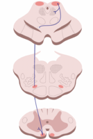
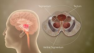
The origin of the Tectospinal tract is in the superior colliculus of the [[midbrain]]. As this area recieves information regarding visual input, this tract is primarily responsible for mediating [[Reflexes|reflex]] responses to visual stimuli. <ref name="Crossman">Crossman AR, Neary D. Neuroanatomy: An Illustrated Colour Text. Third Edition. London: Elsevier, 2004</ref> The tectospinal tract is named after the tectum, meaning roof. The tectum can be interpreted as the 'roof' of the fourth ventrical. The fourth ventricle is made up of the superior and inferior colliculi.<ref name="Carpenter">Carpenter R, Reddi B. Neurophysiology. A conceptual Approach. Fifth Edition. Hodder Arnold:London, 2012.</ref> | |||
[[ | Part of the [[Extrapyramidal and Pyramidal Tracts|Extrapyramidal]] system, see link. | ||
== Anatomy == | == Anatomy == | ||
[[File:Mid-Brain Different Parts.jpeg|thumb|Midbrain Tectum]] | |||
'''Origin:''' Superior colliculus of the midbrain (tectum of the [[midbrain]]).<ref name="Crossman" /><ref name="Fitzgerald">Fitzgerald MJT, Gruener G, Mtui E. Clinical neuroanatomy and neuroscience. Fifth Edition. Philadelphia: Elsevier Saunders, 2007</ref> | |||
'''Course:''' Passes ventromedially around the periaqueductal grey matter. Terminates in the medial part of the anterior gray horm of cervical and upper thoracic segments within laminae VI–VII. <ref name="Crossman" /><ref name="Fitzgerald" /><ref name="Rea" /> | |||
== Function | == Function == | ||
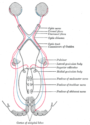
[[Image:Superior colliculus.png|alt=|thumb|Superior Colliculus Midbrain ]]The Tectospinal tract receives information from the retina and cortical visual association areas <ref name="Rea">Rea P. Essential clinical anatomy of the nervous system. Academic Press; 2015 Jan 5.</ref>. In response to visual stimuli, the tectospinal tract mediates reflex movements<ref name="Crossman" />.It is able to orientate the head/trunk towards auditory stimulus (inferior colliculus) or visual stimuli (superior colliculus).<ref name="Fitzgerald" /> | |||
During visual stimuli being sensed the [[Neurone|neurons]] in the superior colliculus respond and cause the eye to saccade to the same part of the visual field. <ref name="Carpenter" /> | |||
Efferent fibres are thereby also sent to the [[Reticular Formation|reticular formation]] that trigger saccades and also spinal regions innervating the [[Cervical Anatomy|neck]] <ref name="Carpenter" />The connections with effector muscles are excitatory to contralateral neck muscle [[Motor Neurone|motor neurons]] and inhibitory to ipsilateral motor neurones.<ref name="Sengull">Sengul1 G, Watson C, Spinal Cord :Connections. In Mai JK, Paxinos G. The Human Nervous System. Third Edition. Academic Press, 2004</ref> | |||
<br> | To discover more about these reflexes see [[Vestibular Anatomy and Neurophysiology]]<br> | ||
== References == | == References == | ||
<references />. | <references />. | ||
Latest revision as of 04:50, 27 April 2022
Original Editor - Kate Sampson
Top Contributors - Kate Sampson, Lucinda hampton, WikiSysop and Kim Jackson
Description[edit | edit source]
The origin of the Tectospinal tract is in the superior colliculus of the midbrain. As this area recieves information regarding visual input, this tract is primarily responsible for mediating reflex responses to visual stimuli. [1] The tectospinal tract is named after the tectum, meaning roof. The tectum can be interpreted as the 'roof' of the fourth ventrical. The fourth ventricle is made up of the superior and inferior colliculi.[2]
Part of the Extrapyramidal system, see link.
Anatomy[edit | edit source]
Origin: Superior colliculus of the midbrain (tectum of the midbrain).[1][3]
Course: Passes ventromedially around the periaqueductal grey matter. Terminates in the medial part of the anterior gray horm of cervical and upper thoracic segments within laminae VI–VII. [1][3][4]
Function[edit | edit source]
The Tectospinal tract receives information from the retina and cortical visual association areas [4]. In response to visual stimuli, the tectospinal tract mediates reflex movements[1].It is able to orientate the head/trunk towards auditory stimulus (inferior colliculus) or visual stimuli (superior colliculus).[3]
During visual stimuli being sensed the neurons in the superior colliculus respond and cause the eye to saccade to the same part of the visual field. [2]
Efferent fibres are thereby also sent to the reticular formation that trigger saccades and also spinal regions innervating the neck [2]The connections with effector muscles are excitatory to contralateral neck muscle motor neurons and inhibitory to ipsilateral motor neurones.[5]
To discover more about these reflexes see Vestibular Anatomy and Neurophysiology
References[edit | edit source]
- ↑ 1.0 1.1 1.2 1.3 Crossman AR, Neary D. Neuroanatomy: An Illustrated Colour Text. Third Edition. London: Elsevier, 2004
- ↑ 2.0 2.1 2.2 Carpenter R, Reddi B. Neurophysiology. A conceptual Approach. Fifth Edition. Hodder Arnold:London, 2012.
- ↑ 3.0 3.1 3.2 Fitzgerald MJT, Gruener G, Mtui E. Clinical neuroanatomy and neuroscience. Fifth Edition. Philadelphia: Elsevier Saunders, 2007
- ↑ 4.0 4.1 Rea P. Essential clinical anatomy of the nervous system. Academic Press; 2015 Jan 5.
- ↑ Sengul1 G, Watson C, Spinal Cord :Connections. In Mai JK, Paxinos G. The Human Nervous System. Third Edition. Academic Press, 2004
.


