Thomas Test: Difference between revisions
No edit summary |
Kim Jackson (talk | contribs) No edit summary |
||
| (83 intermediate revisions by 18 users not shown) | |||
| Line 1: | Line 1: | ||
<div class="editorbox"> | <div class="editorbox"> | ||
'''Original Editor '''- [[User:Tyler Shultz|Tyler Shultz | '''Original Editor '''- [[User:Tyler Shultz|Tyler Shultz]] | ||
'''Top Contributors''' - {{Special:Contributors/{{FULLPAGENAME}}}} | '''Top Contributors''' - {{Special:Contributors/{{FULLPAGENAME}}}} | ||
'''Edited April 2023''' - by [[User:Maddison Jones|Maddison Jones]], [[User:Ashtyn Madden|Ashtyn Madden]], and [[User:Shelby Morrison|Shelby Morrison]] as part of the [[Arkansas Colleges of Health Education School of Physical Therapy Musculoskeletal 1 Project]]</div> | |||
== Purpose == | |||
The Thomas Test measures hip flexor length and distinguishes tightness between one joint and two joint muscles. Hip flexor length directly correlates to the available range of motion at the hip and knee joints.<ref name=":1">Florence Peterson Kendall, McCreary E, Provance P, Rodgers M, Romani W. Muscles : Testing and Function with Posture and Pain. 5th ed. Baltimore, Md: Lippincott Williams & Wilkins; 2005.</ref> | |||
== | Impaired range of motion of the hip may be an underlying cause to other conditions such as: psoas syndrome; [[Patellofemoral Pain Syndrome|patellofemoral pain syndrome]]; [[Low Back Pain|lower back pain]]; [[osteoarthritis]]; [[Rheumatoid Arthritis|rheumatoid arthritis]]. | ||
[[File:Muscles of the hip and thigh - Kenhub.png|frameless|600x600px|Overview of the hip and thigh - anterior and posterior views|center]] | |||
== Relevant Anatomy == | |||
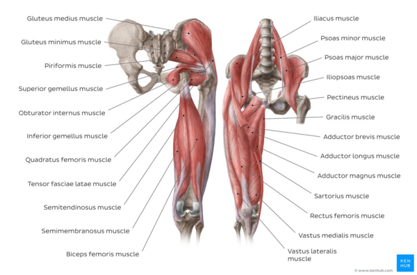
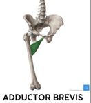
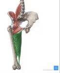
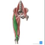
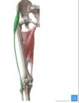
Various muscles are tested during the Thomas test including: | |||
{| class="wikitable" | |||
|'''Hip Flexor Muscle''' | |||
|'''Picture''' | |||
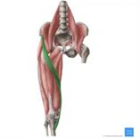
'''(Muscle in Green)'''<ref>Osika A. Hip and thigh muscles [Internet]. Kenhub. 2022. Available from: <nowiki>https://www.kenhub.com/en/library/anatomy/hip-and-thigh-muscles</nowiki></ref> | |||
|'''Number of Joints Crossed''' | |||
|'''Main Function''' | |||
|'''Additional Movement''' | |||
|- | |||
|Iliopsoas: Composed of iliacus and psoas major | |||
|[[File:Iliopsoas Muscle.jpg|center|180x180px|alt=|frameless]] | |||
|One joint: Hip | |||
|Hip flexion | |||
|Hip external rotation | |||
|- | |||
|Pectineus | |||
|[[File:Pectineus Muscle.jpg|center|150x150px|alt=|frameless]] | |||
|One joint: Hip | |||
|Hip flexion | |||
|Hip abduction | |||
|- | |||
|Adductor Longus & Brevis | |||
|[[File:Adductor Longus Muscle.jpg|center|150x150px|alt=|frameless]][[File:Adductor Brevis Muscle.jpg|center|150x150px|alt=|frameless]] | |||
|One joint: Hip | |||
|Hip adduction | |||
|Hip flexion and external rotation | |||
|- | |||
|Adductor Magnus | |||
|[[File:Adductor Magnus Muscle.jpg|center|150x150px|alt=|frameless]] | |||
|One joint: Hip | |||
|Adductor part: Hip flexion, adduction, external rotation | |||
|Hamstring part: Hip extension and internal rotation | |||
|- | |||
|Rectus femoris | |||
|[[File:Rectus Femoris Muscle.jpg|center|153x153px|alt=|frameless]] | |||
|Two joints: Hip and knee | |||
|Hip flexion | |||
|Knee extension | |||
|- | |||
|Tensor fascia lata | |||
|[[File:Tensor Fascia Lata.jpg|center|150x150px|alt=|frameless]] | |||
|Two joints: Hip and knee | |||
|Hip abduction, flexion, internal rotation | |||
|Knee extension | |||
|- | |||
|Sartorius | |||
|[[File:Sartorius Muscle.jpg|center|157x157px|alt=|frameless]] | |||
|Two joints: Hip and knee | |||
|Hip flexion, abduction, external rotation | |||
|Knee flexion | |||
|} | |||
This 16 minute video is a good summary of the lower extremity muscles. | |||
How to Remember Every Muscle of the Lower Limb and Leg | [https://youtu.be/mlnq-HjWRbA Corporis]<ref>How to Remember Every Muscle of the Lower Limb and Leg video- Corporis <nowiki>https://youtu.be/mlnq-HjWRbA</nowiki></ref> | |||
{{#ev:youtube|mlnq-HjWRbA}} | |||
== Equipment == | |||
The following equipment is needed to perform the Thomas test: | |||
* Stable/firm table. | |||
* Goniometer. | |||
* Chart for recording findings.<ref name=":1" /> | |||
== Technique == | |||
'''Starting Position:''' Patient is standing at the end of a table with their gluteal folds on the edge (the same starting position ensures consistency amongst additional tests). The examiner places one hand behind the patient’s knee and another behind their back before helping them to lay back on the table with their knee flexed. The clinician will apply a posterosuperior stabilization force to the ASIS on the side being tested. This stabilization force ensures the pelvis maintains a neutral position. If the pelvis is not stabilized and moves into an anterior tilt, the hip flexors might appear to have an appropriate length, giving a false negative. The patient will keep the unaffected leg flexed, and slowly lower the affected leg, letting it extend as far as possible.<ref name=":1" /> | |||
'''Correct Testing:''' | |||
* Examiner helps to lay the patient onto the table | |||
* Low back and sacrum are flat on the table | |||
* The non-test leg is in 90 degrees of hip flexion (perpendicular to the table) | |||
** Helps to improve accuracy across tests/retests | |||
'''Errors in Testing:''' | |||
* Patient lays back on their own- unless doing the modified test | |||
* Patient pulls hip into too much flexion, creating a posterior tilt of the pelvis which pulls the thigh off table | |||
* Low back and sacrum are not flat on the table | |||
* Bringing both hips into flexion- allows excessive posterior tilt of the pelvis | |||
* Improper pelvic stabilization- allows pelvic anterior tilt, giving the appearance of normal hip flexor length<ref name=":1" /> | |||
'''Modified Version:''' The patient is positioned sitting at the end of an examination table. The patient is then asked to lie down while bringing both knees to their chest. They should then perform a posterior pelvic tilt- flat back. One limb should then be lowered towards the table while keeping the opposite tucked towards their chest.<ref>Dutton M. Dutton’s orthopedic survival guide managing common conditions /. New York, N.Y.: Mcgraw-Hill Education Llc., C; 2011.</ref> | |||
== Viewing == | |||
The following videos demonstrate how to perform the Thomas test and the modified version of the test:{{#ev:youtube|0lxHQiUtSR8}} | |||
{{#ev:youtube|dm5PfU5_aWY}} | |||
== Interpretation == | |||
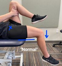
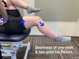
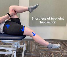
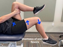
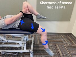
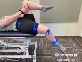
The table below describes various presentations of a positive Thomas test and the muscles affected: | |||
<nowiki>*</nowiki>Images: X- stabilization of ASIS, arrows- direction of movement | |||
{| class="wikitable" | |||
|'''Presentation''' | |||
|'''Muscle(s) Affected''' | |||
|'''Signs''' | |||
|- | |- | ||
| | |Typical length of hip flexors (negative test)[[File:Normal length of hip flexors.jpg|center|215x215px|alt=|frameless]] | ||
| | |Psoas major and iliacus, rectus femoris, tensor fasciae latae, sartorius, pectineus, adductor longus/brevis/magnus | ||
| | |Posterior thigh touches the table | ||
Knee flexes ~ 80° | |||
|- | |- | ||
| | |Shortness of both one-joint and two-joint hip flexors (positive test)[[File:Shortness of one & two-joint muscles.jpg|center|267x267px|alt=|frameless]] | ||
| thigh | |Psoas major and iliacus, rectus femoris, tensor fasciae latae, sartorius, pectineus, adductor longus/brevis/magnus | ||
|Posterior thigh does not touch the table | |||
Knee extends | |||
|- | |- | ||
| | |Shortness of two-joint hip flexors. (positive test)[[File:Two-joint hip flexor tightness.jpg|center|236x236px|alt=|frameless]] | ||
| thigh | |Rectus femoris, tensor fasciae latae, sartorius | ||
|Posterior thigh touches the table | |||
Knee extends | |||
|- | |- | ||
| | |Shortness of one-joint hip flexors. (positive test)[[File:One-joint hip flexor tightness.jpg|center|269x269px|alt=|frameless]] | ||
| thigh | |Iliopsoas, pectineus, adductor longus/brevis/magnus | ||
|Posterior thigh does not touch table | |||
Knee flexes >80° | |||
|- | |- | ||
| | |Shortness of tensor fasciae latae. (positive test)[[File:Tightness of tensor fasciae latae.jpg|center|267x267px|alt=|frameless]] | ||
| | |Tensor fasciae latae | ||
| knee | |Abduction of the thigh as hip joint extends, lateral deviation of the patella. Knee extension if abduction/adduction is prevented with hip extension. Internal rotation of the thigh and external rotation of the leg on the femur. | ||
|- | |||
|Shortness of sartorius. (positive test)[[File:Tightness of Sartorius.jpg|center|267x267px|alt=|frameless]] | |||
|Sartorius | |||
|Abduction, flexion, external rotation of the hip and flexion of the knee. Combination of three or more indicates tightness. | |||
|} | |} | ||
A muscle's strength and available length directly correlate to the range of motion at a joint. Two principles describe the relationship between muscle strength, length, and joint range of motion: | |||
< | '''Active insufficiency:''' Occurs when a two joint muscle produces movement at both joints simultaneously and reaches a shortened length where it can no longer generate force.<ref name=":0">O’Connell A, Gardner E. Understanding the Scientific Basis of Human Motion. Baltimore: Williams & Wilkins; 1972.</ref> <ref name=":4">Kendall F, McCreary E, Provance P. Muscles: Testing and Function with Posture and Pain. 4th ed. Baltimore: Lippincott, Williams & Wilkins; 1993.</ref> Active insufficiency refers to a lack of muscle strength.<ref name=":1" /> | ||
== | '''Passive insufficiency:''' Occurs when a two-joint muscle is in such a lengthened position that it cannot sufficiently permit motion at both joints.<ref name=":0" /> <ref name=":4" /> Passive insufficiency refers to a lack of muscle length, i.e. the muscle is tight.<ref name=":1" /> | ||
Active and passive insufficiency principles describe potential reasons for decreased range of motion at a joint. | |||
== Technique | == '''Goniometry''' == | ||
Goniometric measurements can assist the clinician in determining the available range of motion at a joint. These goniometric measurements are unique to the individual; therefore, they should be compared bilaterally. | |||
The following are considered to be typical goniometric measurements for the hip and knee: | |||
<nowiki>*</nowiki>A- axis, SA- stationary arm, MA- movement arm | |||
{| class="wikitable" | |||
|'''Action''' | |||
|'''Typical Degrees of Motion''' | |||
|'''Technique'''<ref name=":1" /> | |||
|'''Picture''' | |||
|- | |||
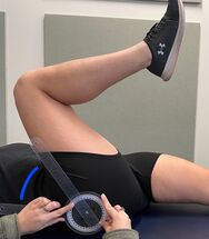
|Hip Flexion | |||
|125°<ref name=":1" /> | |||
|Supine with knee flexed | |||
A: Greater trochanter | |||
SA: Aligned with midline of pelvis | |||
MA: Aligned with femur (lateral epicondyle) | |||
|[[File:Hip Flexion ROM.jpg|alt=|center|215x215px|frameless]] | |||
|- | |||
|Hip Extension | |||
|10°<ref name=":1" /> | |||
|Prone with pelvis stabilized | |||
A: Greater trochanter | |||
SA: Aligned with midline of pelvis | |||
MA: Aligned with femur (lateral epicondyle) | |||
|[[File:Hip Extension ROM.jpg|alt=|center|323x323px|frameless]] | |||
|- | |||
|Hip Internal Rotation | |||
|32°<ref name=":3">Roach KE, Miles TP. Normal Hip and Knee Active Range of Motion: The Relationship to Age. Physical Therapy [Internet]. 1991 Sep 1 [cited 2019 Mar 24];71(9):656–65. Available from: <nowiki>https://pdfs.semanticscholar.org/7e60/2a134667ff5f6f5dda8c7608a59f204d662c.pdf</nowiki></ref> | |||
|Seated on table with knee flexed | |||
A: Center of patella | |||
SA: Aligned perpendicular to the floor | |||
MA: Aligned with leg (crest of tibia) | |||
|[[File:Hip IR.jpg|center|frameless|215x215px]] | |||
|- | |||
|Hip External Rotation | |||
|32°<ref name=":3" /> | |||
|Seated on table with knee flexed | |||
A: Center of patella | |||
SA: Aligned perpendicular to the floor | |||
MA: Aligned with leg (crest of tibia) | |||
|[[File:Hip ER.jpg|center|frameless|215x215px]] | |||
|- | |||
|Knee Flexion | |||
|140°<ref name=":1" /> | |||
|Supine | |||
A: Lateral epicondyle | |||
SA: Greater trochanter | |||
MA: Lateral malleolus | |||
|[[File:Knee Flexion ROM.jpg|alt=|center|215x215px|frameless]] | |||
|- | |||
|Knee Extension | |||
|0°<ref name=":1" /> | |||
|Supine with prop under heel | |||
A: Lateral epicondyle | |||
= | SA: Greater trochanter | ||
MA: Lateral malleolus | |||
|[[File:Knee Extension ROM.jpg|alt=|center|251x251px|frameless]] | |||
|} | |||
== Contraindications == | |||
Contraindications describe conditions that if present suggest the Thomas test should not be performed. Screening for contraindications is a clinical reasoning responsibility of the evaluating physical therapist. | |||
Absolute contraindications: | |||
* Posterior hip replacement | |||
* Suspected fracture- lower extremity, hip, sacrum | |||
Relative contraindications: | |||
* Acute lumbar instability | |||
* Suspected muscle injury- hip flexors | |||
== Reliability == | |||
Studies that test the reliability of the Thomas study are very limited. | |||
== | |||
< | # One study demonstrated that the modified Thomas test has a very good inter-rater reliability.<ref>Gabbe BJ, Bennell KL, Wajswelner H, Finch CF. [https://www.sciencedirect.com/science/article/abs/pii/S1466853X04000227 Reliability of common lower extremity musculoskeletal screening tests.] Physical Therapy in Sport 2004;5(2):90-7.</ref> Another demonstrated that the modified Thomas test, has an average of only moderate levels of reliability.<ref name=":2">Clapis PA, Davis SM, Davis RO. [https://www.tandfonline.com/doi/abs/10.1080/09593980701378256 Reliability of inclinometer and goniometric measurements of hip extension flexibility using the modified Thomas test.] Physiotherapy theory and practice 2008;24(2):135-41.</ref> Further research is required to determine the reliability of the Thomas test. | ||
# Peeler & Anderson conducted a study in 2006 examining the reliability of the Thomas test for assessing [[hip]] range of motion. Their study calls into question the reliability of the technique using both goniometer and pass/fail scoring methods.<ref>Peeler J, Anderson JE. [https://www.sciencedirect.com/science/article/abs/pii/S1466853X06001404 Reliability of the Thomas test for assessing range of motion about the hip.] Physical Therapy in Sport. 2007;8(1):14-21.</ref> | |||
# Boone et al. revealed higher inter-tester reliability for upper-extremity measurements than for lower-extremity measurements, meaning different examiners are more consistent in measuring upper-extremity than lower-extremity range of motion.<ref>Boone DC, Azen SP, Lin CM, Spence C, Baron C, Lee L. Reliability of Goniometric Measurements. Physical Therapy. 1978 Nov 1;58(11):1355–60.</ref><ref name=":3" /> However, other studies have demonstrated very high intra-rater reliability, meaning the same examiner can consistently obtain similar ROM values.<ref>Watkins MA, Riddle DL, Lamb RL, Personius WJ. Reliability of Goniometric Measurements and Visual Estimates of Knee Range of Motion Obtained in a Clinical Setting. Physical Therapy. 1991 Feb 1;71(2):90–6.</ref> | |||
[[Category: | == References == | ||
<references /> | |||
[[Category:Musculoskeletal/Orthopaedics]] | |||
[[Category:Hip]] | |||
[[Category:Sports Medicine]] | |||
[[Category:Athlete Assessment]] | |||
[[Category:Assessment]] | |||
[[Category:Hip - Assessment and Examination]] | |||
[[Category:Muscle Length Testing]] | |||
Latest revision as of 10:43, 18 August 2023
Original Editor - Tyler Shultz
Top Contributors - Aurelie Canas Perez, Maddison Jones, Ashtyn Madden, Admin, Kim Jackson, Magdalena Hytros, Shelby Morrison, Joao Costa, Rachael Lowe, Lucinda hampton, Johnathan Fahrner, Yvonne Yap, Tyler Shultz, Kevin Campion, Kai A. Sigel, Leana Louw, Sue Safadi, Tony Varela, Matt Huey, Oyemi Sillo, Naomi O'Reilly, Adam Vallely Farrell and Wanda van Niekerk
Edited April 2023 - by Maddison Jones, Ashtyn Madden, and Shelby Morrison as part of the Arkansas Colleges of Health Education School of Physical Therapy Musculoskeletal 1 ProjectPurpose[edit | edit source]
The Thomas Test measures hip flexor length and distinguishes tightness between one joint and two joint muscles. Hip flexor length directly correlates to the available range of motion at the hip and knee joints.[1]
Impaired range of motion of the hip may be an underlying cause to other conditions such as: psoas syndrome; patellofemoral pain syndrome; lower back pain; osteoarthritis; rheumatoid arthritis.
Relevant Anatomy[edit | edit source]
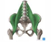
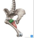
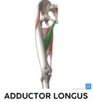
Various muscles are tested during the Thomas test including:
| Hip Flexor Muscle | Picture
(Muscle in Green)[2] |
Number of Joints Crossed | Main Function | Additional Movement |
| Iliopsoas: Composed of iliacus and psoas major | One joint: Hip | Hip flexion | Hip external rotation | |
| Pectineus | One joint: Hip | Hip flexion | Hip abduction | |
| Adductor Longus & Brevis | One joint: Hip | Hip adduction | Hip flexion and external rotation | |
| Adductor Magnus | One joint: Hip | Adductor part: Hip flexion, adduction, external rotation | Hamstring part: Hip extension and internal rotation | |
| Rectus femoris | Two joints: Hip and knee | Hip flexion | Knee extension | |
| Tensor fascia lata | Two joints: Hip and knee | Hip abduction, flexion, internal rotation | Knee extension | |
| Sartorius | Two joints: Hip and knee | Hip flexion, abduction, external rotation | Knee flexion |
This 16 minute video is a good summary of the lower extremity muscles.
How to Remember Every Muscle of the Lower Limb and Leg | Corporis[3]
Equipment[edit | edit source]
The following equipment is needed to perform the Thomas test:
- Stable/firm table.
- Goniometer.
- Chart for recording findings.[1]
Technique[edit | edit source]
Starting Position: Patient is standing at the end of a table with their gluteal folds on the edge (the same starting position ensures consistency amongst additional tests). The examiner places one hand behind the patient’s knee and another behind their back before helping them to lay back on the table with their knee flexed. The clinician will apply a posterosuperior stabilization force to the ASIS on the side being tested. This stabilization force ensures the pelvis maintains a neutral position. If the pelvis is not stabilized and moves into an anterior tilt, the hip flexors might appear to have an appropriate length, giving a false negative. The patient will keep the unaffected leg flexed, and slowly lower the affected leg, letting it extend as far as possible.[1]
Correct Testing:
- Examiner helps to lay the patient onto the table
- Low back and sacrum are flat on the table
- The non-test leg is in 90 degrees of hip flexion (perpendicular to the table)
- Helps to improve accuracy across tests/retests
Errors in Testing:
- Patient lays back on their own- unless doing the modified test
- Patient pulls hip into too much flexion, creating a posterior tilt of the pelvis which pulls the thigh off table
- Low back and sacrum are not flat on the table
- Bringing both hips into flexion- allows excessive posterior tilt of the pelvis
- Improper pelvic stabilization- allows pelvic anterior tilt, giving the appearance of normal hip flexor length[1]
Modified Version: The patient is positioned sitting at the end of an examination table. The patient is then asked to lie down while bringing both knees to their chest. They should then perform a posterior pelvic tilt- flat back. One limb should then be lowered towards the table while keeping the opposite tucked towards their chest.[4]
Viewing[edit | edit source]
The following videos demonstrate how to perform the Thomas test and the modified version of the test:
Interpretation[edit | edit source]
The table below describes various presentations of a positive Thomas test and the muscles affected:
*Images: X- stabilization of ASIS, arrows- direction of movement
| Presentation | Muscle(s) Affected | Signs |
| Typical length of hip flexors (negative test) | Psoas major and iliacus, rectus femoris, tensor fasciae latae, sartorius, pectineus, adductor longus/brevis/magnus | Posterior thigh touches the table
Knee flexes ~ 80° |
| Shortness of both one-joint and two-joint hip flexors (positive test) | Psoas major and iliacus, rectus femoris, tensor fasciae latae, sartorius, pectineus, adductor longus/brevis/magnus | Posterior thigh does not touch the table
Knee extends |
| Shortness of two-joint hip flexors. (positive test) | Rectus femoris, tensor fasciae latae, sartorius | Posterior thigh touches the table
Knee extends |
| Shortness of one-joint hip flexors. (positive test) | Iliopsoas, pectineus, adductor longus/brevis/magnus | Posterior thigh does not touch table
Knee flexes >80° |
| Shortness of tensor fasciae latae. (positive test) | Tensor fasciae latae | Abduction of the thigh as hip joint extends, lateral deviation of the patella. Knee extension if abduction/adduction is prevented with hip extension. Internal rotation of the thigh and external rotation of the leg on the femur. |
| Shortness of sartorius. (positive test) | Sartorius | Abduction, flexion, external rotation of the hip and flexion of the knee. Combination of three or more indicates tightness. |
A muscle's strength and available length directly correlate to the range of motion at a joint. Two principles describe the relationship between muscle strength, length, and joint range of motion:
Active insufficiency: Occurs when a two joint muscle produces movement at both joints simultaneously and reaches a shortened length where it can no longer generate force.[5] [6] Active insufficiency refers to a lack of muscle strength.[1]
Passive insufficiency: Occurs when a two-joint muscle is in such a lengthened position that it cannot sufficiently permit motion at both joints.[5] [6] Passive insufficiency refers to a lack of muscle length, i.e. the muscle is tight.[1]
Active and passive insufficiency principles describe potential reasons for decreased range of motion at a joint.
Goniometry[edit | edit source]
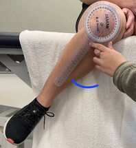
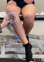
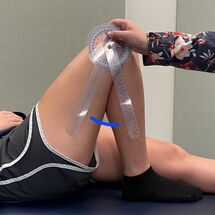
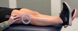
Goniometric measurements can assist the clinician in determining the available range of motion at a joint. These goniometric measurements are unique to the individual; therefore, they should be compared bilaterally.
The following are considered to be typical goniometric measurements for the hip and knee:
*A- axis, SA- stationary arm, MA- movement arm
| Action | Typical Degrees of Motion | Technique[1] | Picture |
| Hip Flexion | 125°[1] | Supine with knee flexed
A: Greater trochanter SA: Aligned with midline of pelvis MA: Aligned with femur (lateral epicondyle) |
|
| Hip Extension | 10°[1] | Prone with pelvis stabilized
A: Greater trochanter SA: Aligned with midline of pelvis MA: Aligned with femur (lateral epicondyle) |
|
| Hip Internal Rotation | 32°[7] | Seated on table with knee flexed
A: Center of patella SA: Aligned perpendicular to the floor MA: Aligned with leg (crest of tibia) |
|
| Hip External Rotation | 32°[7] | Seated on table with knee flexed
A: Center of patella SA: Aligned perpendicular to the floor MA: Aligned with leg (crest of tibia) |
|
| Knee Flexion | 140°[1] | Supine
A: Lateral epicondyle SA: Greater trochanter MA: Lateral malleolus |
|
| Knee Extension | 0°[1] | Supine with prop under heel
A: Lateral epicondyle SA: Greater trochanter MA: Lateral malleolus |
Contraindications[edit | edit source]
Contraindications describe conditions that if present suggest the Thomas test should not be performed. Screening for contraindications is a clinical reasoning responsibility of the evaluating physical therapist.
Absolute contraindications:
- Posterior hip replacement
- Suspected fracture- lower extremity, hip, sacrum
Relative contraindications:
- Acute lumbar instability
- Suspected muscle injury- hip flexors
Reliability[edit | edit source]
Studies that test the reliability of the Thomas study are very limited.
- One study demonstrated that the modified Thomas test has a very good inter-rater reliability.[8] Another demonstrated that the modified Thomas test, has an average of only moderate levels of reliability.[9] Further research is required to determine the reliability of the Thomas test.
- Peeler & Anderson conducted a study in 2006 examining the reliability of the Thomas test for assessing hip range of motion. Their study calls into question the reliability of the technique using both goniometer and pass/fail scoring methods.[10]
- Boone et al. revealed higher inter-tester reliability for upper-extremity measurements than for lower-extremity measurements, meaning different examiners are more consistent in measuring upper-extremity than lower-extremity range of motion.[11][7] However, other studies have demonstrated very high intra-rater reliability, meaning the same examiner can consistently obtain similar ROM values.[12]
References[edit | edit source]
- ↑ 1.00 1.01 1.02 1.03 1.04 1.05 1.06 1.07 1.08 1.09 1.10 Florence Peterson Kendall, McCreary E, Provance P, Rodgers M, Romani W. Muscles : Testing and Function with Posture and Pain. 5th ed. Baltimore, Md: Lippincott Williams & Wilkins; 2005.
- ↑ Osika A. Hip and thigh muscles [Internet]. Kenhub. 2022. Available from: https://www.kenhub.com/en/library/anatomy/hip-and-thigh-muscles
- ↑ How to Remember Every Muscle of the Lower Limb and Leg video- Corporis https://youtu.be/mlnq-HjWRbA
- ↑ Dutton M. Dutton’s orthopedic survival guide managing common conditions /. New York, N.Y.: Mcgraw-Hill Education Llc., C; 2011.
- ↑ 5.0 5.1 O’Connell A, Gardner E. Understanding the Scientific Basis of Human Motion. Baltimore: Williams & Wilkins; 1972.
- ↑ 6.0 6.1 Kendall F, McCreary E, Provance P. Muscles: Testing and Function with Posture and Pain. 4th ed. Baltimore: Lippincott, Williams & Wilkins; 1993.
- ↑ 7.0 7.1 7.2 Roach KE, Miles TP. Normal Hip and Knee Active Range of Motion: The Relationship to Age. Physical Therapy [Internet]. 1991 Sep 1 [cited 2019 Mar 24];71(9):656–65. Available from: https://pdfs.semanticscholar.org/7e60/2a134667ff5f6f5dda8c7608a59f204d662c.pdf
- ↑ Gabbe BJ, Bennell KL, Wajswelner H, Finch CF. Reliability of common lower extremity musculoskeletal screening tests. Physical Therapy in Sport 2004;5(2):90-7.
- ↑ Clapis PA, Davis SM, Davis RO. Reliability of inclinometer and goniometric measurements of hip extension flexibility using the modified Thomas test. Physiotherapy theory and practice 2008;24(2):135-41.
- ↑ Peeler J, Anderson JE. Reliability of the Thomas test for assessing range of motion about the hip. Physical Therapy in Sport. 2007;8(1):14-21.
- ↑ Boone DC, Azen SP, Lin CM, Spence C, Baron C, Lee L. Reliability of Goniometric Measurements. Physical Therapy. 1978 Nov 1;58(11):1355–60.
- ↑ Watkins MA, Riddle DL, Lamb RL, Personius WJ. Reliability of Goniometric Measurements and Visual Estimates of Knee Range of Motion Obtained in a Clinical Setting. Physical Therapy. 1991 Feb 1;71(2):90–6.
