Acetabulum Fracture
Original Editor - Lise Delagrange
Top Contributors - Scott Buxton, 127.0.0.1, Lise Delagrange, Lauren Lopez, Lucinda hampton, Angeliki Chorti, Tony Lowe, Kim Jackson, Candace Goh, Admin, Didzis Rozenbergs, Sweta Christian and WikiSysop
Introduction[edit | edit source]
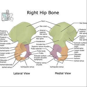
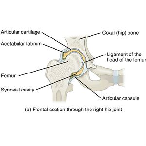
The acetabulum is the large cup-shaped cavity on the anterolateral aspect of the pelvis that articulates with the head of the femur to form the hip joint. Acetabular fractures (a type of pelvic fracture), possibly involving the ilium, ischium or pubis depending on fracture configuration[1].
Epidemiology[edit | edit source]
Acetabular fractures are uncommon. The reported incidence is approximately 3 per 100,000 per year.[1]
Pathology[edit | edit source]
Mechanism
- High-energy trauma: axial loading of the femur: fall from height; Motor vehicle collision; crush injury
- Low-energy trauma with abnormal bone: insufficiency fracture eg Osteoporosis makes the bone vulnerable and can cause an acetabulum fracture from a simple low fall [2].
Diagnostic Procedures[edit | edit source]
There are various numbers of fracture patterns in the acetabulum. For the classification of these fractures, the Judet-Letournel classification is most accepted system. The five most common fractures, representative for 90% of all, are[3]:
- 'Both' column: This is a fracture that involves the anterior and posterior column and extends through the obturator ring. The characteristic for this fracture is the spur sign. It is a sign that represents the displacement of the fracture involving sciatic buttress. When this is present, the acetabulum can no longer carry the weight of the upper body.
- Transverse: This fracture involves both anterior and posterior column. This means that the iliopectineal and ilioischial lines of the pelvis are discontinue.
- T-shaped: Similar to the transverse fractures, the difference is that the T-shaped also extends inferiorly into the obturator ring. What differentiates it from the both column fracture is that the fracture doesn’t involve the extension to the iliac wing.
- 'Transverse' with posterior wall: This is a transverse fracture only there is also a comminution of the posterior wall.
- 'Isolated 'posterior 'wall: One of the most common acetabulum fractures with a preponderance of 27 %. The posterior wall in this fracture is comminuted. The iliopectineal line is not disrupted but the ilioischial line may be [4] [3][5].
Examination[edit | edit source]
CT has revolutionised the diagnosis, enabling precise delineation of the fracture configuration and assessment of any articular surface disruption.[1]
The radiograph gives a clear view of all the essential fundamental landmarks of the acetabulum.
On a CT-scan you can see full 3D- reconstruction of the acetabulum, which facilitates the visualization of the fracture, the degree of the fracture and the associated fractures[6].
Medical Management [edit | edit source]
Treatment depends on several factors including both patient factors and fracture characteristics. Treatment, whether operative or non-operative will usually be followed by a period of non-weight bearing on the affected side (or both). Close radiographic follow-up is required. [1]
If the displacement of the fragment is greater than 3mm, operation is primarily suggested[4]. Non-operative management may be indicated in the setting of minimally displaced fracture. It is more common in developing countries[1].
These fractures often require an open reduction internal fixation (ORIF) to restore joint congruency and stabilization. For the operation they use screws and plates to fix the bone to prevent further displacement[5][7]With elderly patients, ORIF may not always be the best option because of possible osteoporosis. ORIF can be the solution to the fracture if the femoral head is still weight bearing and when the patient factors are no immediate cause to any complication. Patient factors include degree of underlying osteoporosis, comorbid medical conditions, preexisting degenerative joint disease (DJD), premorbid activity level, and baseline mental function[8].
In general total hip replacement will provide the best functional outcome in elderly patients with severe acetabulum fracture and osteoporosis[9].
Physical Management[edit | edit source]
Usually this nonoperative treatment is recommended for patients with no displacements or minimal displacement like low anterior column or low transverse fractures, so the superior part of the acetabulum must be intact[10].
Early mobilization is necessary because prolonged recumbency can be life- threatening[8]. Conservative treatment includes pain control, functional physical therapy and radiographic follow-up[8]. Physical therapy include gait training, stabilization exercises and mobility training.
Patients who underwent an operation have to start with passive ROM exercises followed by active non weight bearing such as a series of flexion/extension . Partial weight-bearing with stepwise progression usually starts 6 weeks postoperatively and full weight bearing is eventually allowed at 10 weeks[11][4].
References[edit | edit source]
- ↑ 1.0 1.1 1.2 1.3 1.4 Radiopedia Acetabular fracture Available:https://radiopaedia.org/articles/acetabular-fracture?lang=gb (accessed 30.10.2022)
- ↑ Kanakaris NK, Davidson A. Classification of acetabular fractures: how to apply and relevance today. Orthopaedics and Trauma. 2022 Apr 1;36(2):61-6.
- ↑ 3.0 3.1 Durkee NJ, Jacobson J, Jamadar D, Karunakar MA, Morag Y, Hayes C. Classification of common acetabular fractures: radiographic and CT appearances. AJR Am J Roentgenol. 2006 Oct 1;187(4):915-25.
- ↑ 4.0 4.1 4.2 Tile M, Helfet DL, Kellam JF, Vrahas M. Fractures of the pelvis and acetabulum third edition. Baltimore: Williams & Wilkins; 2011.
- ↑ 5.0 5.1 Liu X, Xu S, Zhang C, Su J, Yu B. Application of a shape-memory alloy internal fixator for treatment of acetabular fractures with a follow-up of two to nine years in China. International orthopaedics. 2010 Oct;34(7):1033-40.
- ↑ Balendra G, Bassett JW, Acharya M. The ABC management of the acetabular fracture patient. Orthopaedics and Trauma. 2022 Mar 25.
- ↑ Stilger VG, Alt JM, Hubbard DF. Traumatic acetabular fracture in an intercollegiate football player: a case report. Journal of athletic training. 2000 Jan;35(1):103.
- ↑ 8.0 8.1 8.2 Pagenkopf E, Grose A, Partal G, Helfet DL. Acetabular fractures in the elderly: treatment recommendations. HSS Journal®. 2006 Sep;2(2):161-71.
- ↑ Cornell CN. Management of acetabular fractures in the elderly patient. HSS Journal®. 2005 Sep;1(1):25-30.
- ↑ Cochu G, Mabit C, Gougam T, Fiorenza F, Baertich C, Charissoux JL, Arnaud JP. Total hip arthroplasty for treatment of acute acetabular fracture in elderly patients. Revue de chirurgie orthopedique et reparatrice de l'appareil moteur. 2007 Dec 1;93(8):818-27.
- ↑ Kristan A, Mavcic B, Cimerman M, Iglic A, Tonin M, Slivnik T, Kralj-Iglic V, Daniel M. Acetabular loading in active abduction. IEEE Transactions on neural systems and rehabilitation engineering. 2007 Jun 18;15(2):252-7.


