Compartment Syndrome: Difference between revisions
No edit summary |
Kim Jackson (talk | contribs) m (Text replacement - "[[Edema" to "[[Oedema") |
||
| (10 intermediate revisions by 3 users not shown) | |||
| Line 1: | Line 1: | ||
<div class="editorbox"> | <div class="editorbox"> '''Original Editor '''- [[User:Racheal Lowe|Racheal Lowe]] '''Top Contributors''' - {{Special:Contributors/{{FULLPAGENAME}}}}</div> | ||
'''Original Editor ''' | |||
== Introduction == | == Introduction == | ||
[[File:Compartment Syndrome Picture Wikipedia.jpeg|right|frameless|alt=|Compartment syndrome in leg]] | |||
Acute Compartment Syndrome is a condition in which there is increased pressure within a closed osteofascial compartment, resulting in impaired local circulation. Without prompt treatment, acute compartment syndrome can lead to ischemia and eventually, necrosis.<ref name=":2">Torlincasi AM, Lopez RA, Waseem M. [https://www.ncbi.nlm.nih.gov/books/NBK448124/ Acute compartment syndrome].2017 Available: https://www.ncbi.nlm.nih.gov/books/NBK448124/<nowiki/>(accessed 28.10.2021)</ref> | |||
The anterior compartment of the leg is the most common location for compartment syndrome. Other locations in which acute compartment syndrome is seen include the forearm, thigh, buttock, shoulder, hand, and foot.<ref name=":2" /> | |||
See [[Compartment Syndrome of the Lower Leg]]; [[Compartment Syndrome of the Forearm]]; [[Compartment Syndrome of the Foot|Compartment Syndrome of the Foot.]] | |||
== Etiology == | == Etiology == | ||
Acute compartment syndrome can occur without any precipitating trauma but typically occurs after a long bone fracture, with tibial fractures being the most common cause of the condition, followed by distal radius fractures. | Acute compartment syndrome can occur without any precipitating trauma but typically occurs after a long [[Fracture|bone fracture]], with tibial fractures being the most common cause of the condition, followed by [[Distal Radial Fractures|distal radius fractures.]] | ||
* Seventy-five percent of cases of acute compartment syndrome are associated with fractures. After fractures, the most common cause of acute compartment syndrome is soft tissue injuries. | * Seventy-five percent of cases of acute compartment syndrome are associated with fractures. After fractures, the most common cause of acute compartment syndrome is soft tissue injuries. | ||
* Other causes of acute compartment syndrome include burns, vascular injuries, crush injuries, drug overdoses, reperfusion injuries, thrombosis, bleeding disorders, infections, improperly placed casts or splints, tight circumferential bandages, penetrating trauma, intense athletic activity, and poor positioning during surgery. | * Other causes of acute compartment syndrome include [[Burns Overview|burns,]] vascular injuries, crush injuries, [[Substance Use Disorder|drug overdoses]], reperfusion injuries, thrombosis, bleeding disorders, infections, improperly placed casts or [[Splint|splints]], tight circumferential bandages, penetrating trauma, intense athletic activity, and poor positioning during surgery. | ||
* In children, supracondylar fractures of the humerus and both ulnar and radial forearm fractures are associated with compartment syndrome<ref name=":2" />. | * In children, [[Supracondylar Humeral Fracture|supracondylar fractures of the humerus]] and both ulnar and radial forearm fractures are associated with compartment syndrome<ref name=":2" />. | ||
{{#ev:youtube|IOKixPJi-Ns}} | {{#ev:youtube|IOKixPJi-Ns}} | ||
The connective tissue forming a compartment is not pliable, so when bleeding or swelling occurs within the compartment, the intra-compartmental pressure rises.<ref name="bron 5">Kirsten G B, Elliot A, J Johnstone. Diagnosing acute compartment syndrome. The journal of bone and joint surgery, Vol. 85, N°5, July 2003 A1 (2)http://web.jbjs.org.uk/cgi/reprint/85-B/5/625.pdf Level of evidence: A1</ref><ref name="bron 6">Galanakos S, Sakellariou V I, Kkotoulas H, Sofianos I P. Acute Compartment Syndrome: The significance of immediate diagnosis and the consequences from delayed treatment. E.E.X.O.T, Vol 60: 127-133, 2009 Level of evidence: A1</ref> Normally a non-contracting muscle contains a pressure near zero. | == Epidemiology == | ||
The incidence of acute compartment syndrome is estimated to be 7.3 per 100,000 in males and 0.7 per 100,000 in females, with the majority of cases occurring after trauma. Tibial shaft fracture is the most common cause of acute compartment syndrome (associated with a 1 to 10 percent incidence of acute compartment syndrome)<ref name=":2" />. | |||
== Mechanism of Injury / Pathological Process == | |||
The connective tissue forming a compartment is not pliable, so when bleeding or swelling occurs within the compartment, the intra-compartmental pressure rises.<ref name="bron 5">Kirsten G B, Elliot A, J Johnstone. Diagnosing acute compartment syndrome. The journal of bone and joint surgery, Vol. 85, N°5, July 2003 A1 (2)http://web.jbjs.org.uk/cgi/reprint/85-B/5/625.pdf Level of evidence: A1</ref><ref name="bron 6">Galanakos S, Sakellariou V I, Kkotoulas H, Sofianos I P. Acute Compartment Syndrome: The significance of immediate diagnosis and the consequences from delayed treatment. E.E.X.O.T, Vol 60: 127-133, 2009 Level of evidence: A1</ref> | |||
Normally a non-contracting muscle contains a pressure near zero. | |||
* If the pressure rises up to 30 mmHg, the vessels will be compressed, resulting in pain and a decrease in blood flow. | |||
*[[Lymphatic System|Lymphatic]] drainage will activate to prevent increasing interstitial fluid pressure.<ref name="bron 4">Abraham T Rasul Jr. Compartment syndrome. eMedicine. 11 March 2009 A1 (2)http://emedicine.medscape.com/article/307668-overview Level of evidence: A1</ref> | |||
* Once the effects of lymphatic drainage have reached their maximum, the pressure within the compartments will cause physiological defects, such as a nerve dysfunction and deformation. | |||
== Clinical Presentation | * Haemorrhage or [[Oedema Assessment|oedema]] causes the interstitial pressures within the soft tissues to increase, creating possible ischemia by loss of [[Capillary Refill Test|capillary refill]].<ref name="bron 7">Tucker Alicia K. Chronic exertional compartment syndrome of the leg. Current Reviews in Musculoskeletal Medicine. 2 September 2010 A1 http://ukpmc.ac.uk/articles/PMC2941579/ Level of evidence: A1</ref> | ||
* When a body part is not provided with blood for more than eight hours, the damage is irreversible and may lead to the death of the concerning tissues.<ref name="bron 3">Frink M, Hildebrand F, Krettek C, Brand J, Hankemeier S. Compartment syndrome of the lower leg and foot. The Association of bone and joint surgeons. 27 may 2009 http://emedicine.medscape.com/article/140002-overview Level of evidence: B</ref> | |||
== Clinical Presentation == | |||
=== Symptoms of Chronic Compartment Syndrome === | === Symptoms of Chronic Compartment Syndrome === | ||
Obtaining an accurate patient history is vital, due to the objective examination often not showing much of note. In a typical case, the patient will present with pain in a compartment of the leg, at the same time, distance and intensity of exercise.<ref>Cook S, Bruce G. Fasciotomy for chronic compartment syndrome in the lower limb. ANZ J Surg 2002; 72(10):720-3</ref> The pain shall continue to increase until it becomes unbearable and the patient stops exercising, causing the pain to subside with rest. | Obtaining an accurate patient history is vital, due to the objective examination often not showing much of note. In a typical case, the patient will present with [[Pain Assessment|pain]] in a compartment of the leg, at the same time, distance and intensity of exercise.<ref>Cook S, Bruce G. Fasciotomy for chronic compartment syndrome in the lower limb. ANZ J Surg 2002; 72(10):720-3</ref> The pain shall continue to increase until it becomes unbearable and the patient stops exercising, causing the pain to subside with rest. | ||
*Pain on palpation of involved muscles | *Pain on palpation of involved muscles | ||
*Pain with passive stretching of muscles | *Pain with passive [[stretching]] of muscles | ||
*The feeling of firmness of involved compartments | *The feeling of firmness of involved compartments | ||
*Muscle herniation can be palpated in 40-60% of patients with compartment syndrome (Usually palpated over anterior tibia) | *Muscle herniation can be palpated in 40-60% of patients with compartment syndrome (Usually palpated over anterior tibia) | ||
*Gait analysis may show excessive overpronation | *[[Gait|Gait analysis]] may show excessive overpronation | ||
*A neurological exam may show weakness and numbness of the affected compartment<br> | *A neurological exam may show weakness and numbness of the affected compartment<br> | ||
Remember the 5 P’s: Pain, Pallor, Paresthesia, Paralysis, Pulselessnes'''s'''<ref name="bron 4" /> | |||
The | == Prognosis == | ||
The prognosis after treatment of compartment syndrome depends mainly on how quickly the condition is diagnosed and treated. When fasciotomy is done within 6 hours, there is almost 100% recovery of limb function. After 6 hours, there may be residual [[Nerve Injury Rehabilitation|nerve]] damage. Data show that when the fasciotomy is done within 12 hours, only two-thirds of patients have normal limb function. In very delayed cases, the limb may require an [[Amputations|amputation]]. | |||
=== | == Diagnostic Procedures == | ||
# Intra-Compartmental Pressure Monitoring (ICP): Not required but can aid in diagnosis if uncertainty exists. Compartment pressures are often measured with a manometer, a device that detects intracompartmental pressure by measuring the resistance that is present when a saline solution is injected into the compartment. Another method employs a slit catheter, whereby a catheter is placed within the compartment, and the pressure measured with an arterial line transducer. <br> | |||
# Less Invasive Measurement Techniques | |||
#*Laser Doppler ultrasound | |||
#*Methoxy isobutyl isonitrile enhanced magnetic resonance imaging ([[MRI Scans|MRI]]) | |||
#*Phosphate-nuclear magnetic resonance (NMR) spectroscopy | |||
== | == Outcome Measures == | ||
* | *[[Lower Extremity Functional Scale (LEFS)|Lower Extremity Functional Scale]] (LEFS)<ref name=":0">Tjeerdsma J. Outcome of a specific compartment fasciotomy versus a complete compartment fasciotomy of the leg in one patient with bilateral anterior chronic exertional compartment syndrome: a case report. The Journal of Foot and Ankle Surgery. 2016 Sep 1;55(5):1027-34.</ref> | ||
* | *[[Foot and Ankle Ability Measure]] (FAAM)<ref name=":0" /> | ||
* | *[[Visual Analogue Scale]]<ref name=":1">Meulekamp MZ, van der Wurff P, van der Meer A, Lucas C. Identifying prognostic factors for conservative treatment outcomes in servicemen with chronic exertional compartment syndrome treated at a rehabilitation center. Military Medical Research. 2017 Dec;4(1):36.</ref> | ||
*[[Patient Specific Functional Scale]]<ref name=":1" /> | |||
For more see [[Outcome Measures|Outcome Measures Database]] | |||
== | == Management / Interventions == | ||
[[File:1024px-Compartment syndrome with fasciotomy procedure 01.jpeg|right|frameless|355x355px]] | |||
In the event of a diagnosis of Compartment syndrome (when there is a intra-compartment pressure of >30 mmHg<ref name="p1">Von Schroeder HP et al. Definitions and terminology of compartment syndrome and Volkmann's ischemic contracture of the upper extremity. Hand Clin. 1998 Aug;14(3):331-4.</ref><ref name="p2">Jim Clover. Sports medicine essentials, core concepts in athletic training. 2nd edition. 2010</ref>) immediate surgical fasciotomy is needed to reduce the intracompartmental pressure. | |||
Image 2: Compartment syndrome with fasciotomy procedure | |||
* The ideal timeframe for fasciotomy is within six hours of injury | |||
*The | * Fasciotomy is not recommended after 36 hours following injury. When tissue pressure remains elevated for that amount of time, irreversible damage may occur, and fasciotomy may not be beneficial in this situation. | ||
* | |||
After | After a fasciotomy is performed and swelling dissipates, a skin graft is commonly used for incision closure. Patients must be closely monitored for complications which include infection, acute renal failure, and rhabdomyolysis. | ||
If necrosis occurs before fasciotomy is performed, there is a high likelihood of infection which may require amputation. If infection occurs, debridement is necessary to prevent systemic spread or other complications<ref name=":2" />. | |||
Physiotherapy role in the treatment of the condition is vital, with or without surgical intervention. The physiotherapist may employ modalities that will improve range of motion, strength of the affected muscles, function and relief pain. | == Physiotherapy == | ||
Physiotherapy role in the treatment of the condition is vital, with or without surgical intervention. The physiotherapist may employ modalities that will improve range of motion, strength of the affected muscles, function and relief pain. | |||
See highlighted links in Introduction for site specific Physiotherapy | |||
== Differential Diagnosis == | == Differential Diagnosis == | ||
| Line 85: | Line 87: | ||
These common pathologies may give the same pain characteristics or symptoms in the lower limbs:<ref name="bron 20">http://www.physioadvisor.com.au/10513350/compartment-syndrome-chronic-compartment-syndrom.htm</ref> | These common pathologies may give the same pain characteristics or symptoms in the lower limbs:<ref name="bron 20">http://www.physioadvisor.com.au/10513350/compartment-syndrome-chronic-compartment-syndrom.htm</ref> | ||
*[[ | *[[Medial Tibial Stress Syndrome|shin splints]] (medial tibial stress syndrome) | ||
*[[Stress Fractures|stress fractures]] | *[[Stress Fractures|stress fractures]] | ||
*[[Leg and Foot Stress Fractures|fascial defects]] | *[[Leg and Foot Stress Fractures|fascial defects]] | ||
Latest revision as of 11:45, 3 August 2022
Introduction[edit | edit source]
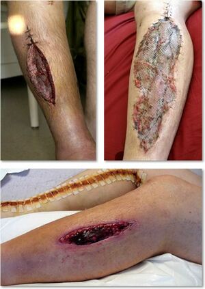
Acute Compartment Syndrome is a condition in which there is increased pressure within a closed osteofascial compartment, resulting in impaired local circulation. Without prompt treatment, acute compartment syndrome can lead to ischemia and eventually, necrosis.[1]
The anterior compartment of the leg is the most common location for compartment syndrome. Other locations in which acute compartment syndrome is seen include the forearm, thigh, buttock, shoulder, hand, and foot.[1]
See Compartment Syndrome of the Lower Leg; Compartment Syndrome of the Forearm; Compartment Syndrome of the Foot.
Etiology[edit | edit source]
Acute compartment syndrome can occur without any precipitating trauma but typically occurs after a long bone fracture, with tibial fractures being the most common cause of the condition, followed by distal radius fractures.
- Seventy-five percent of cases of acute compartment syndrome are associated with fractures. After fractures, the most common cause of acute compartment syndrome is soft tissue injuries.
- Other causes of acute compartment syndrome include burns, vascular injuries, crush injuries, drug overdoses, reperfusion injuries, thrombosis, bleeding disorders, infections, improperly placed casts or splints, tight circumferential bandages, penetrating trauma, intense athletic activity, and poor positioning during surgery.
- In children, supracondylar fractures of the humerus and both ulnar and radial forearm fractures are associated with compartment syndrome[1].
Epidemiology[edit | edit source]
The incidence of acute compartment syndrome is estimated to be 7.3 per 100,000 in males and 0.7 per 100,000 in females, with the majority of cases occurring after trauma. Tibial shaft fracture is the most common cause of acute compartment syndrome (associated with a 1 to 10 percent incidence of acute compartment syndrome)[1].
Mechanism of Injury / Pathological Process[edit | edit source]
The connective tissue forming a compartment is not pliable, so when bleeding or swelling occurs within the compartment, the intra-compartmental pressure rises.[2][3]
Normally a non-contracting muscle contains a pressure near zero.
- If the pressure rises up to 30 mmHg, the vessels will be compressed, resulting in pain and a decrease in blood flow.
- Lymphatic drainage will activate to prevent increasing interstitial fluid pressure.[4]
- Once the effects of lymphatic drainage have reached their maximum, the pressure within the compartments will cause physiological defects, such as a nerve dysfunction and deformation.
- Haemorrhage or oedema causes the interstitial pressures within the soft tissues to increase, creating possible ischemia by loss of capillary refill.[5]
- When a body part is not provided with blood for more than eight hours, the damage is irreversible and may lead to the death of the concerning tissues.[6]
Clinical Presentation[edit | edit source]
Symptoms of Chronic Compartment Syndrome[edit | edit source]
Obtaining an accurate patient history is vital, due to the objective examination often not showing much of note. In a typical case, the patient will present with pain in a compartment of the leg, at the same time, distance and intensity of exercise.[7] The pain shall continue to increase until it becomes unbearable and the patient stops exercising, causing the pain to subside with rest.
- Pain on palpation of involved muscles
- Pain with passive stretching of muscles
- The feeling of firmness of involved compartments
- Muscle herniation can be palpated in 40-60% of patients with compartment syndrome (Usually palpated over anterior tibia)
- Gait analysis may show excessive overpronation
- A neurological exam may show weakness and numbness of the affected compartment
Remember the 5 P’s: Pain, Pallor, Paresthesia, Paralysis, Pulselessness[4]
Prognosis[edit | edit source]
The prognosis after treatment of compartment syndrome depends mainly on how quickly the condition is diagnosed and treated. When fasciotomy is done within 6 hours, there is almost 100% recovery of limb function. After 6 hours, there may be residual nerve damage. Data show that when the fasciotomy is done within 12 hours, only two-thirds of patients have normal limb function. In very delayed cases, the limb may require an amputation.
Diagnostic Procedures[edit | edit source]
- Intra-Compartmental Pressure Monitoring (ICP): Not required but can aid in diagnosis if uncertainty exists. Compartment pressures are often measured with a manometer, a device that detects intracompartmental pressure by measuring the resistance that is present when a saline solution is injected into the compartment. Another method employs a slit catheter, whereby a catheter is placed within the compartment, and the pressure measured with an arterial line transducer.
- Less Invasive Measurement Techniques
- Laser Doppler ultrasound
- Methoxy isobutyl isonitrile enhanced magnetic resonance imaging (MRI)
- Phosphate-nuclear magnetic resonance (NMR) spectroscopy
Outcome Measures[edit | edit source]
- Lower Extremity Functional Scale (LEFS)[8]
- Foot and Ankle Ability Measure (FAAM)[8]
- Visual Analogue Scale[9]
- Patient Specific Functional Scale[9]
For more see Outcome Measures Database
Management / Interventions[edit | edit source]
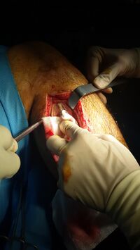
In the event of a diagnosis of Compartment syndrome (when there is a intra-compartment pressure of >30 mmHg[10][11]) immediate surgical fasciotomy is needed to reduce the intracompartmental pressure.
Image 2: Compartment syndrome with fasciotomy procedure
- The ideal timeframe for fasciotomy is within six hours of injury
- Fasciotomy is not recommended after 36 hours following injury. When tissue pressure remains elevated for that amount of time, irreversible damage may occur, and fasciotomy may not be beneficial in this situation.
After a fasciotomy is performed and swelling dissipates, a skin graft is commonly used for incision closure. Patients must be closely monitored for complications which include infection, acute renal failure, and rhabdomyolysis.
If necrosis occurs before fasciotomy is performed, there is a high likelihood of infection which may require amputation. If infection occurs, debridement is necessary to prevent systemic spread or other complications[1].
Physiotherapy[edit | edit source]
Physiotherapy role in the treatment of the condition is vital, with or without surgical intervention. The physiotherapist may employ modalities that will improve range of motion, strength of the affected muscles, function and relief pain.
See highlighted links in Introduction for site specific Physiotherapy
Differential Diagnosis[edit | edit source]
These common pathologies may give the same pain characteristics or symptoms in the lower limbs:[12]
- shin splints (medial tibial stress syndrome)
- stress fractures
- fascial defects
- peroneal nerve entrapment
- popliteal artery entrapment syndrome
- claudication
References[edit | edit source]
- ↑ 1.0 1.1 1.2 1.3 1.4 Torlincasi AM, Lopez RA, Waseem M. Acute compartment syndrome.2017 Available: https://www.ncbi.nlm.nih.gov/books/NBK448124/(accessed 28.10.2021)
- ↑ Kirsten G B, Elliot A, J Johnstone. Diagnosing acute compartment syndrome. The journal of bone and joint surgery, Vol. 85, N°5, July 2003 A1 (2)http://web.jbjs.org.uk/cgi/reprint/85-B/5/625.pdf Level of evidence: A1
- ↑ Galanakos S, Sakellariou V I, Kkotoulas H, Sofianos I P. Acute Compartment Syndrome: The significance of immediate diagnosis and the consequences from delayed treatment. E.E.X.O.T, Vol 60: 127-133, 2009 Level of evidence: A1
- ↑ 4.0 4.1 Abraham T Rasul Jr. Compartment syndrome. eMedicine. 11 March 2009 A1 (2)http://emedicine.medscape.com/article/307668-overview Level of evidence: A1
- ↑ Tucker Alicia K. Chronic exertional compartment syndrome of the leg. Current Reviews in Musculoskeletal Medicine. 2 September 2010 A1 http://ukpmc.ac.uk/articles/PMC2941579/ Level of evidence: A1
- ↑ Frink M, Hildebrand F, Krettek C, Brand J, Hankemeier S. Compartment syndrome of the lower leg and foot. The Association of bone and joint surgeons. 27 may 2009 http://emedicine.medscape.com/article/140002-overview Level of evidence: B
- ↑ Cook S, Bruce G. Fasciotomy for chronic compartment syndrome in the lower limb. ANZ J Surg 2002; 72(10):720-3
- ↑ 8.0 8.1 Tjeerdsma J. Outcome of a specific compartment fasciotomy versus a complete compartment fasciotomy of the leg in one patient with bilateral anterior chronic exertional compartment syndrome: a case report. The Journal of Foot and Ankle Surgery. 2016 Sep 1;55(5):1027-34.
- ↑ 9.0 9.1 Meulekamp MZ, van der Wurff P, van der Meer A, Lucas C. Identifying prognostic factors for conservative treatment outcomes in servicemen with chronic exertional compartment syndrome treated at a rehabilitation center. Military Medical Research. 2017 Dec;4(1):36.
- ↑ Von Schroeder HP et al. Definitions and terminology of compartment syndrome and Volkmann's ischemic contracture of the upper extremity. Hand Clin. 1998 Aug;14(3):331-4.
- ↑ Jim Clover. Sports medicine essentials, core concepts in athletic training. 2nd edition. 2010
- ↑ http://www.physioadvisor.com.au/10513350/compartment-syndrome-chronic-compartment-syndrom.htm


