Brainstem: Difference between revisions
No edit summary |
No edit summary |
||
| Line 17: | Line 17: | ||
==Brainstem Anatomy and Structure== | ==Brainstem Anatomy and Structure== | ||
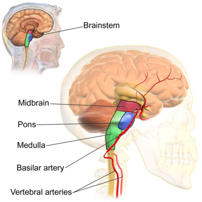
The brainstem is located in the posterior cranial fossa of the skull <ref>Nolte J. The Human Brain: An Introduction to its fuctional Anatomy. St. Louis: Mosby, 1993.</ref>. Recall that the nervous system is a continuous neuronal network. When looking at the brain stem in situ: | [[File:Brain_Stem.jpg|right|frameless|400x400px]]The brainstem is located in the '''posterior cranial fossa''' of the [[skull]] <ref>Nolte J. The Human Brain: An Introduction to its fuctional Anatomy. St. Louis: Mosby, 1993.</ref>. The brainstem can be divided into three regions or components: (1) the midbrain, (2) the pons, and (3) the medulla. | ||
Recall that the nervous system is a continuous neuronal network. When looking at the brain stem in situ: | |||
* superiorly or ventrally , the midbrain is continuous with the [[Cerebrum|cerebral hemisphere]] | * superiorly or ventrally , the midbrain is continuous with the [[Cerebrum|cerebral hemisphere]] | ||
| Line 23: | Line 25: | ||
*posteriorly, the pons and medulla are separated from the [[cerebellum]] by the fourth ventricle | *posteriorly, the pons and medulla are separated from the [[cerebellum]] by the fourth ventricle | ||
=== Midbrain (Mesencephalon) === | |||
The midbrain is the smallest portions of the brainstem and is continuous with the diencephalon (area of the brain which contains epithalamus, thalamus, hypothalamus, and ventral thalamus and the third ventricle). | |||
==== Posterior midbrain: ==== | |||
* the '''tectum''' (i.e. the roof) of the midbrain is formed by two pairs of rounded protrusions: the superior and inferior colliculi: | |||
** '''superior colliculi''' are involved in visual processing and eye movements | |||
** '''inferior colliculi''' are involved in auditory processing | |||
* also connects and communicates with the '''cerebellum''' posteriorly through the '''superior cerebellar peduncles''' | |||
==== Anterior midbrain: ==== | |||
* contains the '''cerebral peduncles''' which are a pair of fibre bundles. separated by the interpeduncular fossa. | |||
** the '''corticospinal tract''', a descending motor tract between the cortex and the spinal cord, is found in the cerebral peduncles | |||
==== Clinically relevant brainstem nuclei found in the midbrain ==== | |||
* '''Red nuclei''', located anterior medially at the level of the superior colliculus. The exact function of the red nuclei is still being investigated, however it is likely involved in locomotor functions and in the execution of "simple, stereotyped movements," and specialized movements of the upper limb.<ref>Basile GA, Quartu M, Bertino S, Serra MP, Boi M, Bramanti A, Anastasi GP, Milardi D, Cacciola A. [https://www.ncbi.nlm.nih.gov/pmc/articles/PMC7817566/ Red nucleus structure and function: from anatomy to clinical neurosciences]. Brain Structure and Function. 2021 Jan;226:69-91.</ref> The red nuclei is the origin of the descending '''rubrospinal tract'''. The rubrospinal tract functions primarily to transmit signals into the red nucleus from the cerebral motor cortex and cerebellum to the spinal cord.<ref>Lee J, Muzio M. Neuroanatomy, Extrapyramidal System [Internet]. 2022 [cited 01/June/2024]. Available from:https://www.ncbi.nlm.nih.gov/books/NBK554542/</ref> | |||
* | * '''[[Substantia Nigra|Substantia nigra]]''', found at the base of the midbrain. The substantia nigra is rich in [[dopamine]] neurons and is considered part of the [[Basal Ganglia|basal ganglia]]. This paired nuclei has a critical role in modulating motor movement and reward functions. In [[Parkinson's Disease|Parkinson's,]] [[Neurodegenerative Disease|neurodegeneration]] occurs due to death of dopamine producing cells in the substantia nigra, which causes the hallmark movement dysfunction seen with this disease process. | ||
* '''Cranial Nerve IV''' (CN IV, the [[Trochlear Nerve|trochlear nerve]]) is found Just inferior to the inferior colliculi. CN IV is unique among its fellow cranial nerves as it ss the only one to emerge from the posterior surface of the brainstem.<ref>Basinger H, Hogg J. Neuroanatomy, Brainstem [Internet]. 2023 [cited 01/June/2024]. Available from: https://www.ncbi.nlm.nih.gov/books/NBK544297/</ref> | |||
* '''Cranial nerve III''' (CN III, the [[Oculomotor Nerve|oculomotor nerve]]) . The oculomotor nerve originates at the oculomotor sulcus within the interpeduncular cistern. | |||
=== Pons (metencephalon) === | |||
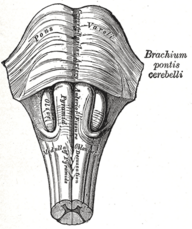
[[File:Pons and medulla oblongata, anterior.png|thumb|Pons and medulla, anterior.|229x229px]] | [[File:Pons and medulla oblongata, anterior.png|thumb|Pons and medulla, anterior.|229x229px]] | ||
The pons is a part of the [[brainstem]] that is located between the [[midbrain]] (cranially) and the medulla oblongata (caudally). It derives its name from its appearance on the anterior surface, which looks like a bridge connecting the right and left cerebellar hemispheres.<ref>Angeles Fernández-Gil M, Palacios-Bote R, Leo-Barahona M, Mora-Encinas JP. [https://www.sciencedirect.com/science/article/abs/pii/S0887217110000260 Anatomy of the brainstem: a gaze into the stem of life.] Seminars in ultrasound, CT, and MR [Internet]. 2010 Jun 1;31(3):196–219.</ref> It also forms important connections with the cerebellum via fibre bundles known as the cerebellar peduncles. | The pons is a part of the [[brainstem]] that is located between the [[midbrain]] (cranially) and the medulla oblongata (caudally). It derives its name from its appearance on the anterior surface, which looks like a bridge connecting the right and left cerebellar hemispheres.<ref>Angeles Fernández-Gil M, Palacios-Bote R, Leo-Barahona M, Mora-Encinas JP. [https://www.sciencedirect.com/science/article/abs/pii/S0887217110000260 Anatomy of the brainstem: a gaze into the stem of life.] Seminars in ultrasound, CT, and MR [Internet]. 2010 Jun 1;31(3):196–219.</ref> It also forms important connections with the cerebellum via fibre bundles known as the cerebellar peduncles. | ||
| Line 43: | Line 55: | ||
*Additionally, cranial nerve nuclei in the pons are involved in several other functions, including swallowing, tear production, hearing, and maintaining [[balance]]/equilibrium. | *Additionally, cranial nerve nuclei in the pons are involved in several other functions, including swallowing, tear production, hearing, and maintaining [[balance]]/equilibrium. | ||
=== Medulla (myelencephalon) === | |||
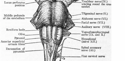
[[File:Pyramidal decussation.png|thumb|407x407px|Shows Pyramidal decussation]] | [[File:Pyramidal decussation.png|thumb|407x407px|Shows Pyramidal decussation]] | ||
The point where the brainstem connects to the [[Spinal cord anatomy|spinal cord]]. | The point where the brainstem connects to the [[Spinal cord anatomy|spinal cord]]. | ||
Revision as of 02:36, 2 June 2024
Original Editor - Wendy Walker
Lead Editors - Lucinda hampton, Olajumoke Ogunleye, Stacy Schiurring, Pacifique Dusabeyezu, Evan Thomas, Kim Jackson, Wendy Walker, Garima Gedamkar, Aya Alhindi, WikiSysop, Rucha Gadgil, Carina Therese Magtibay, Tarina van der Stockt and Daniele Barilla
Brainstem Overview[edit | edit source]
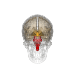
The brainstem is a stalk-like projection of the brain extending caudally from the base of the cerebrum. It function is to bridge communication between the cerebrum with the cerebellum, and spinal cord.[1]
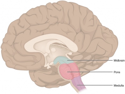
- It is composed of 3 sections in descending order: the midbrain, pons, and medulla oblongata.
- It is responsible for many vital functions of life, such as (1) breathing, (2) consciousness, (3) maintaining blood pressure, (4) heart rate, and (5) sleep regulation.
- The brainstem contains both collections of white and grey matter
- The grey matter within the brainstem consist of nerve cell bodies and include important brainstem nuclei, for example, ten of the twelve cranial nerves arise from their cranial nerve nuclei located within the brainstem.
- The white matter tracts of the brainstem involve neuron axons traveling between the cerebrum, cerebellum, and spinal cord to the peripheral nervous system. Most of the cell bodies of these white matter tracts originate outside the brainstem, however some are located within the brainstem as well. These tracts travel both to the brain (afferent) and from the brain (efferent) such as the somatosensory pathways and the corticospinal tracts, respectively.
- Although it is the most evolutionary ancient part of our brain, the brainstem is still very complex and important. It ensures the vital functions necessary to support those areas continue uninterrupted.[2]
Brainstem Anatomy and Structure[edit | edit source]
The brainstem is located in the posterior cranial fossa of the skull [3]. The brainstem can be divided into three regions or components: (1) the midbrain, (2) the pons, and (3) the medulla.
Recall that the nervous system is a continuous neuronal network. When looking at the brain stem in situ:
- superiorly or ventrally , the midbrain is continuous with the cerebral hemisphere
- inferiorly or caudally, the medulla is continuous with the spinal cord
- posteriorly, the pons and medulla are separated from the cerebellum by the fourth ventricle
Midbrain (Mesencephalon)[edit | edit source]
The midbrain is the smallest portions of the brainstem and is continuous with the diencephalon (area of the brain which contains epithalamus, thalamus, hypothalamus, and ventral thalamus and the third ventricle).
Posterior midbrain:[edit | edit source]
- the tectum (i.e. the roof) of the midbrain is formed by two pairs of rounded protrusions: the superior and inferior colliculi:
- superior colliculi are involved in visual processing and eye movements
- inferior colliculi are involved in auditory processing
- also connects and communicates with the cerebellum posteriorly through the superior cerebellar peduncles
Anterior midbrain:[edit | edit source]
- contains the cerebral peduncles which are a pair of fibre bundles. separated by the interpeduncular fossa.
- the corticospinal tract, a descending motor tract between the cortex and the spinal cord, is found in the cerebral peduncles
Clinically relevant brainstem nuclei found in the midbrain[edit | edit source]
- Red nuclei, located anterior medially at the level of the superior colliculus. The exact function of the red nuclei is still being investigated, however it is likely involved in locomotor functions and in the execution of "simple, stereotyped movements," and specialized movements of the upper limb.[4] The red nuclei is the origin of the descending rubrospinal tract. The rubrospinal tract functions primarily to transmit signals into the red nucleus from the cerebral motor cortex and cerebellum to the spinal cord.[5]
- Substantia nigra, found at the base of the midbrain. The substantia nigra is rich in dopamine neurons and is considered part of the basal ganglia. This paired nuclei has a critical role in modulating motor movement and reward functions. In Parkinson's, neurodegeneration occurs due to death of dopamine producing cells in the substantia nigra, which causes the hallmark movement dysfunction seen with this disease process.
- Cranial Nerve IV (CN IV, the trochlear nerve) is found Just inferior to the inferior colliculi. CN IV is unique among its fellow cranial nerves as it ss the only one to emerge from the posterior surface of the brainstem.[6]
- Cranial nerve III (CN III, the oculomotor nerve) . The oculomotor nerve originates at the oculomotor sulcus within the interpeduncular cistern.
Pons (metencephalon)[edit | edit source]
The pons is a part of the brainstem that is located between the midbrain (cranially) and the medulla oblongata (caudally). It derives its name from its appearance on the anterior surface, which looks like a bridge connecting the right and left cerebellar hemispheres.[7] It also forms important connections with the cerebellum via fibre bundles known as the cerebellar peduncles.
- Home to several nuclei for cranial nerves.
- Nerves that carry information about sensations of touch, pain, and temperature from the face and head synapse in a nucleus in the pons.
- Motor commands dealing with eye movement, chewing, and facial expressions also originate in the pons.
- Additionally, cranial nerve nuclei in the pons are involved in several other functions, including swallowing, tear production, hearing, and maintaining balance/equilibrium.
Medulla (myelencephalon)[edit | edit source]
The point where the brainstem connects to the spinal cord.
- Contains a nucleus called the nucleus of the solitary tract that is crucial for our survival (receives information about blood flow, along with information about levels of oxygen and carbon dioxide in the blood, from the heart and major blood vessels). When this information suggests a discordance with bodily needs (e.g. blood pressure is too low), there are reflexive actions initiated in the nucleus of the solitary tract to bring things back to within the desired range.
- Essential to our survival because it ensures vital systems e.g cardiovascular and respiratory systems are working properly.
- Responsible for several reflexive actions, including vomiting, swallowing, coughing, and sneezing. Several cranial nerves also exit the brainstem at the level of the medulla.
- Pyramidal decussation is the crossing of the fibers of the corticospinal tracts from one side of the central nervous system to the other near the junction of the medulla and the spinal cord
NB. "Bulb" is an archaic term for the medulla oblongata, the word bulbar (e.g. bulbar palsy) is retained for terms that relate to the medulla oblongata. The word bulbar can refer to the nerves and tracts connected to the medulla, and also by association to the muscles thus innervated, such as those of the tongue, pharynx and larynx.[8]
Blood supply of the Brainstem[edit | edit source]
The blood supply into the brain comes from the vertebral and internal carotid arteries. The vertebral arteries supply the brain stem directly. The paired vertebral arteries come together on the anterior brainstem to form the basilar artery, which lies in the basilar groove on the pons. The network formed by the vertebral and basilar arteries is called the vertebral-basilar system.
- The midbrain is supplied by the superior cerebellar arteries and posterior cerebral arteries
- The pons is supplied by the basilar artery and its branches. This includes the anterior inferior cerebellar arteries, the pontine branches or paramedian arteries, and superior cerebellar arteries
- The medulla oblongata is supplied by the vertebral arteries and their branches, the anterior spinal artery, the posterior spinal arteries, and the posterior inferior cerebellar arteries
Function[edit | edit source]
The brainstem has three broad functions:
1. Serves as a conduit for the ascending tracts and descending tracts connecting the spinal cord to the different parts of the higher centres in the forebrain.
2. Contains important reflex centres associated with the control of:
- respiration e.g: Automatic breathing[9]
- cardiovascular system e.g BP
- consciousness
- autonomic functions such as digestion, salivation, perspiration, dilation or contraction of the pupils, urination, etc.
3. Contains the nuclei of Cranial Nerves III to XII[2].
Clinical Significance[edit | edit source]
Significant clinical problems can affect the brainstem such as stroke, malignancy, demyelinating processes, and many more. e.g
- Multiple Sclerosis, with visual problems including blurred double vision being a common early symptom of MS.
- Stroke affecting the brainstem can cause severe symptoms which include:
- Problems with vital functions, such as breathing - frequently resulting in death.
- Difficulty using with chewing, swallowing, and speaking.
- Weakness or paralysis in the arms, legs, and/or face.
- Problems with balance or sensation.
- Hearing loss
- Vision problems
- Vertigo
- Locked-in Syndrome
- Coma [10]
- Hemiballismus from damage to the subthalamic nucleus.
- Injury to or degeneration of dopaminergic neurons in substantia nigra resulting in Parkinson’s disease.
- Wallenberg stroke (spinothalamic tract, spinal trigeminal tract, hypothalamospinal tract, vestibular nuclei).
- Cerebellar tonsillar herniation (sudden respiratory and cardiac arrest due to compression of the medulla).
- Medial pontine syndrome (abducens nerve, corticospinal tract, medial lemniscus).
- Central pontine myelinolysis from the rapid correction of hyponatremia, which can result in seizures, ataxia, and disturbed consciousness.
The 9 minute video below gives a good summary of brainstem and stroke[11]
References[edit | edit source]
- ↑ Brainstem. Kenhub.
- ↑ 2.0 2.1 Neuroscientifically challenged: Know your brain [Internet]. 2014 [cited 9th January 2021].
- ↑ Nolte J. The Human Brain: An Introduction to its fuctional Anatomy. St. Louis: Mosby, 1993.
- ↑ Basile GA, Quartu M, Bertino S, Serra MP, Boi M, Bramanti A, Anastasi GP, Milardi D, Cacciola A. Red nucleus structure and function: from anatomy to clinical neurosciences. Brain Structure and Function. 2021 Jan;226:69-91.
- ↑ Lee J, Muzio M. Neuroanatomy, Extrapyramidal System [Internet]. 2022 [cited 01/June/2024]. Available from:https://www.ncbi.nlm.nih.gov/books/NBK554542/
- ↑ Basinger H, Hogg J. Neuroanatomy, Brainstem [Internet]. 2023 [cited 01/June/2024]. Available from: https://www.ncbi.nlm.nih.gov/books/NBK544297/
- ↑ Angeles Fernández-Gil M, Palacios-Bote R, Leo-Barahona M, Mora-Encinas JP. Anatomy of the brainstem: a gaze into the stem of life. Seminars in ultrasound, CT, and MR [Internet]. 2010 Jun 1;31(3):196–219.
- ↑ World Heritage Encyclopedia. Medulla Oblongata. http://www.ebooklibrary.org/Articles/Medulla%20oblongata?&Words=Medulla (Accessed 8 April 2017).
- ↑ Herrero JL, Khuvis S, Yeagle E, Cerf M, Mehta AD. Breathing above the brain stem: volitional control and attentional modulation in humans. Journal of Neurophysiology. 2018 Jan 1;119(1):145–59.
- ↑ Brain Injury Explanation [Internet]. [accessed 8 April 2017]. Available from: http://www.braininjury-explanation.com/consequences/impact-by-brain-area/brainstem
- ↑ Basinger H, Hogg JP. Neuroanatomy, Brainstem. StatPearls [Internet]. 2020 [cited 9 January 2021]. Available from:. https://www.ncbi.nlm.nih.gov/books/NBK544297/
- ↑ Soton brain hub Brainstem Stroke Syndromes Available from: https://www.youtube.com/watch?v=qxiRfP9XmpE (last accessed 28.11.2019)
