Flexion Deformity of the Knee
Original Editor - Jentel Van De Gucht
Top Contributors - Laura Ritchie, Jenis Bhalavat, Jentel Van De Gucht, Kim Jackson, Salma Ashraf, Shreya Pavaskar, Shaimaa Eldib, Rachael Lowe, Gayatri Jadav Upadhyay, Saeed Dokhnan, Vidya Acharya, Khloud Shreif, 127.0.0.1, Sehriban Ozmen, Evan Thomas, Scott Buxton and WikiSysop
Definition/Description[edit | edit source]
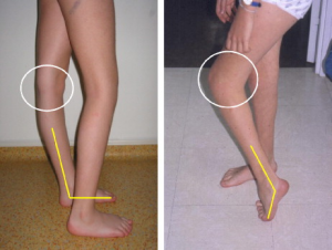
A flexion deformity of the knee is the inability to fully straighten or extend the knee, also known as flexion contracture. Normal active range of motion (AROM) of the knee is 0° extension and 140° flexion. An accurate definition of this would be limited knee extension range[1], both actively and passively. It develops as a result of failure of knee flexors i.e Hamstring muscle to lengthen in tandem with the bone, especially when there is inadequate physical therapy to provide active and passive mobilization of the affected joint.[2] It is usually a combination of bony deformity, capsular and ligamentous deformity. They often require extensive rehabilitation.[3] In most cases, flexion deformities occur bilaterally. The deformity is either temporary or permanent.
| [4] |
Anatomy[edit | edit source]
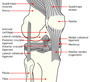
Normal Knee anatomy is characterized by the following:
- Muscle balance: quadriceps versus hamstrings
- Straight leg raise >60º
- Popliteal angle (from horizontal) >60º
- Sagittal plane: full extension
- Straight line between femoral cortex and tibial cortex (see the image below)
- Open physes
- Patella location: between the Blumensat line and physis
- Ground reaction force passes anterior to knee's center of rotation; knee locks passively in extension
- Posterior capsule, gastrocnemius, and hamstrings resist recurvatum.
Epidemiology/Aetiology[edit | edit source]
Flexion deformities can arise by different causes. Two types of flexion contracture of the knee can be distinguished
1) Contracture associated with joint destruction and ankylosis, like,
- Rheumatoid arthritis
- Osteoarthritis
- Cerebral Palsy or congenital deformity - hamstring spasticity
- Hip joint injuries
- Ankle Joint pathologies
- Other degenerative conditions
- Osteogenesis Imperfecta
- Pterygium Syndrome
- Poliomyelitis[5]
2) Contractures with joint anatomy and mobility are preserved:[6]
- After knee operations(Total Knee arthroplasty)
- Tendon transfers
- Stiffness post fractures of Femur, Tibia, Patella or the whole knee joint
- Scar tissue[7]
Pathophysiology[edit | edit source]
Normal sagittal alignment includes the ability to lock the knee in full extension, stabilized posteriorly by the cruciate ligaments , posterior capsule, Hamstrings, and Gastrocnemius. This permits the child to bear full weight without pain, instability, or fatigue, because the ground reaction force is slightly anterior to the extended knee, allowing the child to lock the knee in extension during stance.
- In Cerebral Palsy and Associated Conditions, spastic Hip flexors and Hamstrings combine to flex the knee, causing the ground reaction force to pass behind it and produce a flexion moment. With compromise of the hip extensors and Quadriceps Muscle, gravity and fatigue force the child into a progressive crouch gait pattern[8][9][10] Knee pain is a frequent complaint, which may reflect fatigue of the Quadriceps Muscle, tension failure of the patellar ligament or its bony attachments, or both. Ankle Joint or hindfoot valgus will contribute to lever arm insufficiency and further decrease the extensor moment at the knee.
- Children with Spina Bifida often have intrinsic weakness of the quadriceps, combined with sparing or (if tethered) spasticity of the hamstrings. This puts them at risk for the same problem of FKFD and progressive crouch gait[11] Compounded by ankle valgus, and perhaps planovalgus of the foot, they too have lever arm dysfunction with loss of push-off strength.
- In osteoarthritis or rheumatoid arthritis, swelling is due to synovial inflammation leading to fluid in joint subsequently resulting in assuming of position maximum accommodation i.e. flexion. Chronic posterior femoral and tibial osteophytes tent upon the capsule resulting in further flexion at the knee and sometimes mechanical block to extension.
- Other factors like hamstring shortening and Ligament Contractures also contribute to flexion at the knee. This can lead to increased abnormal forces at the joint while standing, walking, etc and thus lead to abnormal gait pattern which can further lead to limb length discrepancy. Due to these forces and compensatory action of the body to walk, pathological changes may start ascending upwards towards the Pelvis and spine and worsen the condition in severe flexion deformities of knee[12].It is usually associated with either Genu varus or Genu valgus [13].
Characteristics/Clinical Presentation[edit | edit source]
Patients with flexion contractures often walk with a bent-knee gait. Patients often report sleeping with a pillow under their knee or in the fetal position. All of these activities exacerbate the flexion contracture. This provides increasing strain on the Quadriceps Muscle and increasing strain contact forces in the patellofemoral joint and Tibiofemoral Joint when the flexion deformity is more than 15 degress of extensor lag.
There is early joint degradation that includes Cartilage erosion, Meniscal Lesions, Ligament Sprain, associated tightness of TFL and the main muscles around the hip and ankle joint like iliopsoas, Hamstrings, Gastrosoleus, Quadriceps and adductors or abductors of hip depending upon if there is a secondary deformity of either genu varum or genu valgum and patella alta.
Grades of flexion deformity by Lombardi et al[14] -
Grade I - mild contracture with deformity limited to less than 15°
Grade II - moderate contracture with deformity between 15° and 30°
Grade III - severe contracture with deformity greater than 30°
Gait Changes:[edit | edit source]
- Flexed position of the knee at the initiation of the stance phase and throughout the gait cycle. Heel strike is absent, the foot is placed flat on the floor when contracture less than 15 degrees of extensor lag[17] and toe walking where contracture more than 15 degrees of extensor lag. The popliteal angle is reduced.
- The body is propelled forward with increased flexion at hip in swing phase
- A progressive crouch gait and limping while walking leads to shortening of stride length,[18]
- Other symptoms of flexion contractures are anterior knee pain, compensatory movements such as hip flexion deformity accompanied by lumbar lordosis. [16]
- Changes which appear later are severe contracture of knee and hip and patella alta.[19] Knee flexion contracture significantly influences three-dimensional trunk kinematics during relaxed standing and level walking[20], and will lead to spinal imbalance. Due to continuous pressure on the popliteal fossa there may be pressure generated on the common peroneal nerve and tibial nerve and the other contents of fossa[21].
Knee flexion contractures have a lot of functional consequences such as weight-bearing activities and difficulties with bed or chair positioning. Normal daily activities become more difficult because more energy is required to perform them. It interferes with the patient's personal and social life.
In CP, for individuals who are ambulatory, Gross Motor Function Classification System (GMFCS) I–III, limited ability for full knee extension can lead to significant disability with a flexed knee gait posture called crouch gait[22].
History and physical examination[edit | edit source]
In the ambulatory patient with knee flexion deformity , an obligatory crouch gait will be obvious, but it is not necessarily symmetrical. In the seated position, patella alta may be evident, along with reduced power of voluntary knee extension. The femoral condyles may be prominent with an empty sulcus, reflecting the proximal migration of the patella. There may be prominence and tenderness at either pole of the patella, over the tibial tuberosity, or both. Whereas there may be knee crepitance, an effusion is generally not present, because of the chronic nature of the problem.
In the supine position, a straight leg raise should be evaluated. If the degree of knee flexion increases as the hip is flexed (increased popliteal angle), then a concomitant hamstring contracture is likely. If there is no change in the popliteal angle with limb elevation (a bent leg raise), then FKD is the diagnosis[23].
In the prone position, a torsional profile should be documented, as well as the inward-outward range, including hip rotation, and the thigh-foot axis. While the patient is prone, it is easy to look for dynamic versus fixed hip flexion deformity and rectus femoris contracture (Ely test). Also, with the hamstrings relaxed, one can recognize FKD because the ankle/foot will not rest be resting on the table.
Special Tests[edit | edit source]
- Thomas Test: Rule out iliopsoas tightness
- Tripod sign: Hamstring Contracture
- Clarke's test: Patellofemoral pain syndrome
Physiotherapy Management:[edit | edit source]
Depending on etiology and severity of the deformity, different management programs are necessary. Treatment of knee flexion contractures includes non-surgical and surgical methods. [3] In both cases, physiotherapy is necessary. Conservative treatments include physical therapy, home exercise programs, and home mechanical therapy. These are used to treat and minimize the occurrence of flexion contractures.[15] In some cases, such as with cerebral palsy, spasticity management is also necessary. [16]Another method that can help to straighten a knee is use of a device called an extensionator.
The main aim of the treatment is:[24]
- Co-activation of Hamstrings and Quadriceps
- Improve eccentric hamstring strength
- Improve concentric quadriceps strength
- Patellar mobility
- Hip and ankle joint movements
- Gait training
- Return to normal life.
Physical therapy may include manual stretching, prolonged stretching using a tilt table, prolonged stretching using a sandbag/weight over the distal femur, mechanical traction, passive range of motion exercises [25][3] and joint mobilization [3] The effectiveness of a given treatment to reduce flexion contractures is a function of the applied torque, as well as the duration and frequency of the treatment. [15]
Medical Management[edit | edit source]
For patients who have failed standard conservative treatment for two or more months, focused treatment protocols including physical therapy and the use of custom knee devices have been demonstrated to effectively treat flexion contractures. [15] Other treatment methods include orthoses, casting and bracing.[6][3][16] Some types of splits have been marketed as another method of applying low stretching forces over prolonged periods. They provide resistance to flexion so the knee is at rest in maximum extension. The resistance can be inflated. They are easy to apply, mobile and comfortable for patients. [2] In most cases, splints and orthoses are used to prevent deformities or maintain range of motion after stretching but not for increasing motion. [3]
In more severe cases, surgical treatment such as soft-tissue release, osteotomies (removing a part of the bone), femoral shortening, hamstring lengthening and rectus transfer may be necessary.[6][19] Hamstring lengthening is helpful to relieve excessive contractures, especially when they have a significant effect on gait. Rectus transfer may be indicated to partially reduce the spasticity of the quadriceps, especially in patients with cerebral palsy. [28][19]
In spite of all the surgical efforts and post-operative rehabilitation strategies, the deformity can recur and lead to persistent flexion contracture. These patients need manipulation under anaesthesia to get rid of the deformity.[29]
There are specific situations in which the best option is gradual distraction and extension employing an external fixator (Illizarov). This may best serve those patients who have neglected or teratologic deformities, such as pterygium syndrome, or have reached skeletal maturity.
Contraindications for surgery -
There are few contraindications for surgical correction of FKFD (Flexed Knee Flexion Deformity).
Contraindications for osteotomy include the following:
- Nonambulatory status
- Stiff or unstable knee
- Severe osteopenia (fixation issues)
- Flaccid paraplegia
Contraindications for guided growth include the following:
- Closed physes
- Stiff or unstable knee
| [30] |
References:[edit | edit source]
- ↑ Khatri K, Bansal D, Rajpal K. Management of Flexion Contracture in Total Knee Arthroplasty. InKnee Surgery-Reconstruction and Replacement 2020 Apr 22. IntechOpen.
- ↑ 2.0 2.1 Kwan MK, Treatment for flexion contracture of the knee during Ilizarov reconstruction of tibia with passive knee extension splint, 2004;59:39-41 (C)
- ↑ 3.0 3.1 3.2 3.3 3.4 3.5 Suksathien R., A new static progressive splint for treatment of knee and elbow flexion contractures, 2010; 93 (7): 799-804 (C)
- ↑ OrthoMed Pain & Sports Medicine. Flexion Contracture KNEE. Available from: http://www.youtube.com/watch?v=aqifMmasQiM [last accessed 28/08/16]
- ↑ Patricia M. de Moraes Barros Fucs,corresponding author1,2 Celso Svartman,1 and Rodrigo Montezuma,Knee flexion deformity from Poliomyelitis treated by supracondylar femoral extension osteotomy,NCBI,PMCID: PMC2231572
- ↑ 6.0 6.1 6.2 damsin JP, Treatment of severe flexion deformity of the knee in children and adolescents using the Ilizarov technique, 1995;77-B:140-4 (C)
- ↑ Viktor M Grishkevich1* and Vishnevsky AV2,Postburn Knee Flexions Contractures: Anatomy and Methods of Their Treatment,omicsonline.org,October 07, 2013,
- ↑ Arnold AS, Anderson FC, Pandy MG, Delp SL. Muscular contributions to hip and knee extension during the single limb stance phase of normal gait: a framework for investigating the causes of crouch gait. J Biomech. 2005 Nov. 38 (11):2181-9.
- ↑ Delp SL, Arnold AS, Piazza SJ. Graphics-based modeling and analysis of gait abnormalities. Biomed Mater Eng. 1998. 8 (3-4):227-40.
- ↑ Wren TA, Rethlefsen S, Kay RM. Prevalence of specific gait abnormalities in children with cerebral palsy: influence of cerebral palsy subtype, age, and previous surgery. J Pediatr Orthop. 2005 Jan-Feb. 25 (1):79-83.
- ↑ Arnold AS, Anderson FC, Pandy MG, Delp SL. Muscular contributions to hip and knee extension during the single limb stance phase of normal gait: a framework for investigating the causes of crouch gait. J Biomech. 2005 Nov. 38 (11):2181-9.
- ↑ Khatri K, Bansal D, Rajpal K. Management of Flexion Contracture in Total Knee Arthroplasty. InKnee Surgery-Reconstruction and Replacement 2020 Apr 22. IntechOpen.
- ↑ Su EP. Fixed flexion deformity and total knee arthroplasty. The Journal of bone and joint surgery. British volume. 2012 Nov;94(11_Supple_A):112-5.
- ↑ Lombardi Jr AV, Mallory TH, Adams JB, Herrington SM. A stepwise algorithmic approach to flexion contractures in total knee arthroplasty. Arch Am Acad Orthop Surg. 1997;1:1-8.
- ↑ 15.0 15.1 15.2 15.3 Timothy L., Torque Measures of Common Therapies for the Treatment of Flexion Contractures 2010; 26:328-334 (D)
- ↑ 16.0 16.1 16.2 16.3 Klatt J. Guided Growth for Fixed Knee Flexion Deformity, 2008;28:626-631 (C)
- ↑ walking with bend knees,Available from:https://www.youtube.com/watch?v=4rD8MN2fB9o,[Last accessed:Nov 16,2012]
- ↑ Joseph Jankovic, Alberto Albanese, M. Zouhair Atassi, J. Oliver Dolly, Mark Hallett, Nathaniel H. Mayer,Botulinum Toxin E-Book: Therapeutic Clinical Practice and Science,Philadelphia,Saunders Elsevier, pg 197,
- ↑ 19.0 19.1 19.2 Wheeless' Textbook of Orthopaedics (secondary)
- ↑ Harato K1, Nagura T, Matsumoto H, Otani T, Toyama Y, Suda Y.A gait analysis of simulated knee flexion contracture to elucidate knee-spine syndrome.Gait Posture. 2008 Nov;28(4):687-92. doi: 10.1016/j.gaitpost.2008.05.008. Epub 2008 Jun 26.
- ↑ imaios.com,popliteal fossa
- ↑ Lerner ZF, Damiano DL, Bulea TC. A lower-extremity exoskeleton improves knee extension in children with crouch gait from cerebral palsy. Science translational medicine. 2017 Aug 23;9(404).
- ↑ Thompson NS, Baker RJ, Cosgrove AP, Saunders JL, Taylor TC. Relevance of the popliteal angle to hamstring length in cerebral palsy crouch gait. J Pediatr Orthop. 2001 May-Jun. 21 (3):383-7.
- ↑ By HSS, JeMe Cioppa-Mosca, Janet B. Cahill, Carmen Young Tucker,Postsurgical Rehabilitation Guidelines for the Orthopedic Clinician - E-Book pg 273,USA,MOSBY Elsevier,
- ↑ steffen T., Low-Load, Prolonged Stretch in the Treatment of Knee Flexion Contractures in Nursing Home Residents, 1995; 75886-897.1 (A2)
- ↑ hutchjj1. Rotational mobilizations for achieving knee extension. Available from: http://www.youtube.com/watch?v=bhRTEJObwxc [last accessed 28/08/16]
- ↑ MikeReinold.com. The Best and Easiest Way to Restore Knee Extension. Available from: http://www.youtube.com/watch?v=Ui7XKzbXgr8 [last accessed 06/08/16]
- ↑ Mauro C, Treatment of fixed knee flexion deformity and crouch gait using distal femur extension osteotomy in cerebral palsy, 2008, 2(1): 37–43 (C)
- ↑ Sancheti P, Gugale S, Shyam A. Total Knee Arthroplasty in Knees with Fixed Flexion Deformity. InKnee Arthroplasty 2022 (pp. 47-57). Springer, Singapore.
- ↑ Mahkotaorthopaedics. Guided Growth for Fixed Knee Flexion Deformity. Available from: http://www.youtube.com/watch?v=b_j327371fM [last accessed 28/08/16]
|}


